Radiology-Pathology Correlation on Core Biopsy
Contents
- 1 Overview
- 2 Quick Reference - Rad/Path Correlation Chart
- 3 Quick Reference - What To Do Next
- 4 Calcifications
- 5 Background Information - Commonly Used Radiological Terms
- 6 Background Information - Imaging Modalities
- 7 Background Information - Breast Imaging Reporting and Data System (BI-RADS)
- 8 Background Information - Types Of Core Biopsies
- 9 Background Information - Specimen Radiography
| Fine linear calcifications seen on imaging often correlate with low-grade DCIS. | 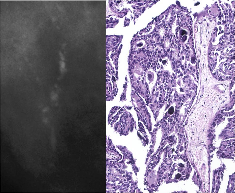 |
Overview
Breast imaging is used for screening and for investigation of areas of clinical concern. The detection of an indeterminate or suspicious lesion usually leads to a biopsy. The radiologist and the pathologist must co‑operate to ensure that the histologic findings explain the radiographic findings; otherwise, the lesion might have been inadequately sampled or might be incompletely examined.
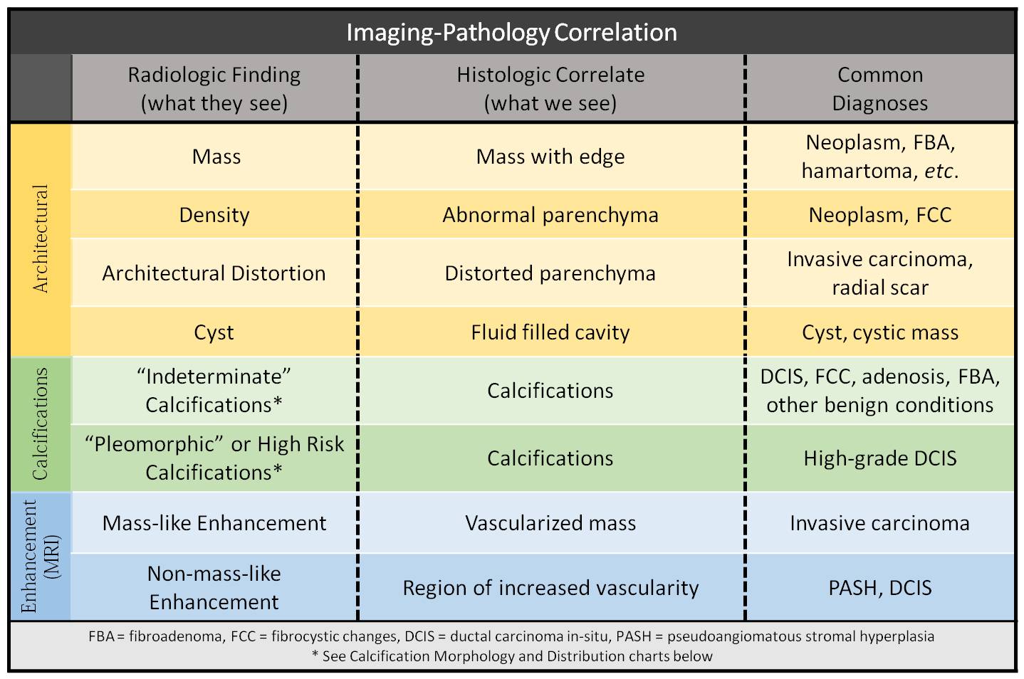
For pathologists, correlation begins with consideration of the radiographic findings. Calcifications, masses, and signal abnormalities are the most common indications for biopsy and each has its histologic correlates. For example, finding an area of dense parenchyma may explain a mass, but it is incidental in the search for calcifications.
Refer to the following chart when correlating your histological findings with the radiological findings.
Quick Reference - Rad/Path Correlation Chart
 |
|
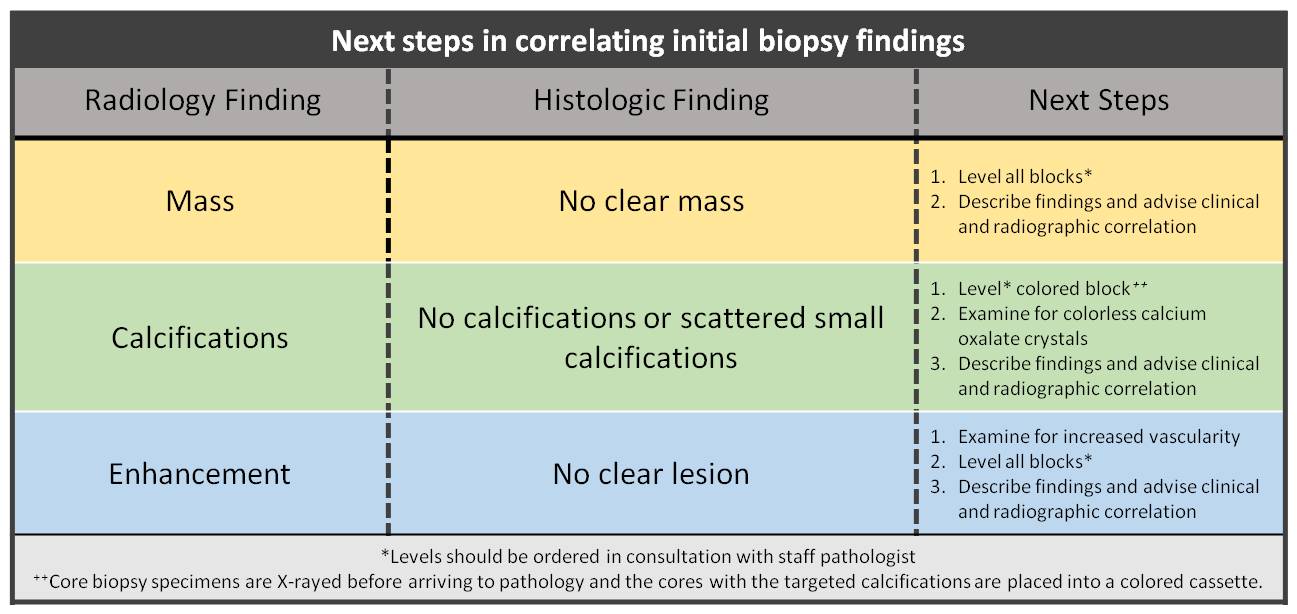
Quick Reference - What To Do Next
 |
|
Calcifications
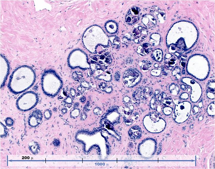
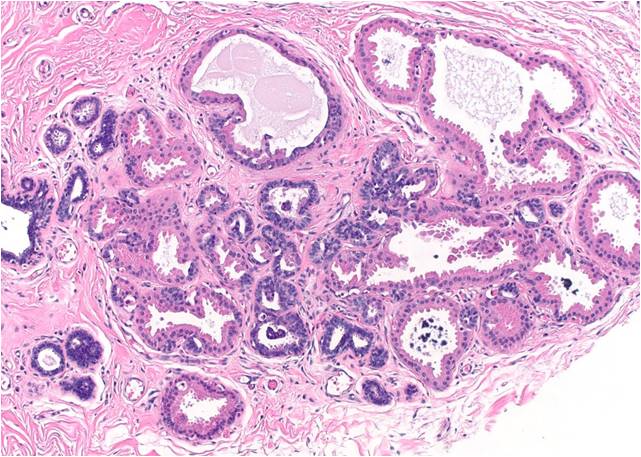
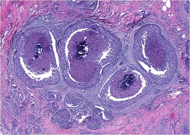
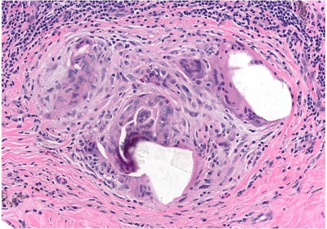
Calcifications are extremely common and their frequency increases with age. In fact, calcifications of one type or another are seen on more than 75% of mammograms. Radiologists separate calcifications into benign, intermediate, and highly suspicious categories based on their morphology and distribution.
 |
|
Morphology and Distribution
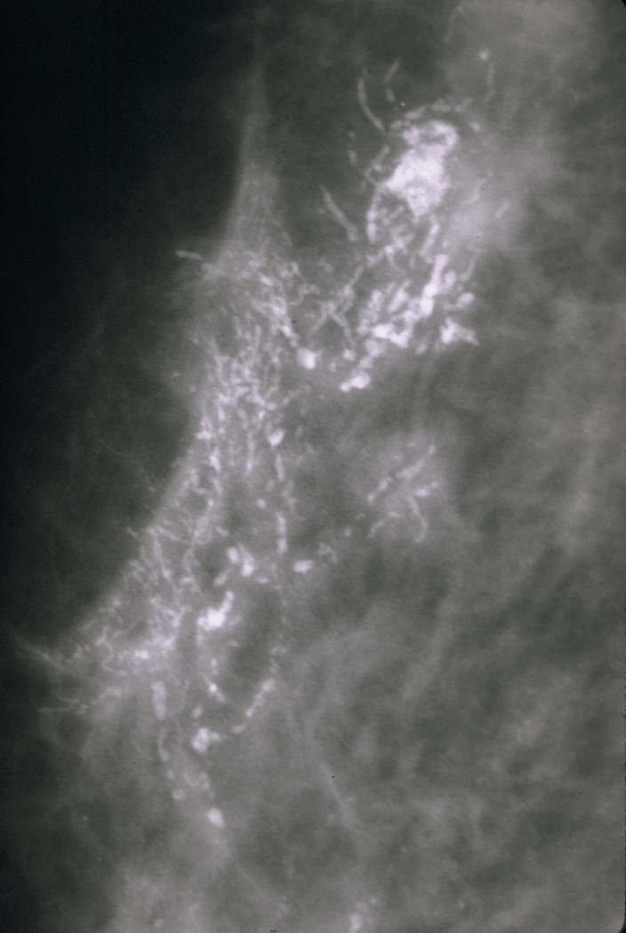
- The morphology and distribution of calcifications depends in part on whether they form in lobules or in ducts. Those that form in lobules usually originate from benign processes that fill and dilate acini in a broad distribution. As a result, lobular calcifications tend to have large, sharply outlined morphologies and to be distributed throughout the breast or in a large region of the breast. Calcifications that have a classical lobular morphology and distribution are classified as benign. Calcifications formed in duct lumens are usually derived from cellular debris or secretions. Like the materials that they arise from, these calcifications tend to be pleomorphic in size and shape and to be distributed in linear or branching patterns. Calcifications of a ductal type are suspicious for malignancy.
- Additionally, calcifications can form in association with the other constituents of the breast, such as the skin, fat, and blood vessels. When they are easily identified as such, these calcifications are classified as benign and do not provoke a biopsy.
Composition of Calcifications
|
 |
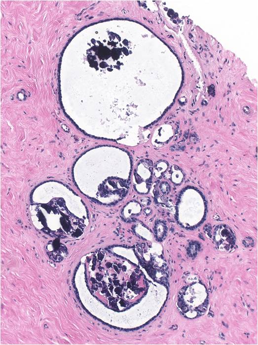 |
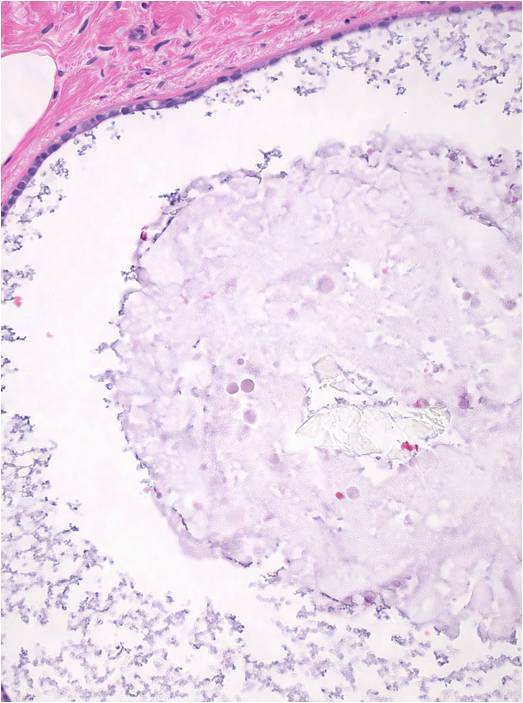 |
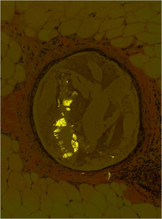 |
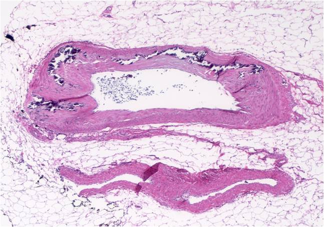
Calcification Size Experiments have shown that calcifications smaller than 0.2 mm (200 microns) are unlikely to be detected radiographically. Since calcifications smaller than 200 microns are easily detected microscopically, it is important to keep in mind this limitation and to ensure that the calcifications encountered histologically are large enough to account for the calcifications that prompted biopsy. If the histologic calcifications are smaller than 200 microns (either singly or in tight aggregates), then it is essential to examine deeper levels or X-ray the blocks to ensure correlation. Also, since the radiographically observed calcifications may be much larger than the 200 micron minimum, it is always prudent to review the radiographs.
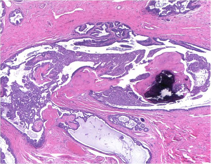 |
 |
 |
 |
 |
 |
Background Information - Commonly Used Radiological Terms
- Mass - Space occupying lesion seen in two different projections (three dimensional).
- Density - Potential mass seen in only a single projection (called a mass if its three-dimensional nature is confirmed).
Architectural Distortion - The normal architecture is distorted, but a mass is not visible. This includes spiculations radiating from a point and focal retraction or distortion of a parenchyma edge.
- Non-masslike Enhancement - Area of enhancement on MRI without three-dimensional characteristics. Non-masslike enhancements are associated with a high risk of malignancy.
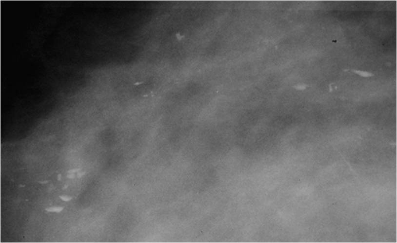
Milk of Calcium - Calcification in the form of a liquid slurry which falls dependently to the bottom of a cyst. Because the material is liquid, it runs out of the tissue during staining.
- Tomosynthesis - Screening imaging methodology that produces CT-like images at a very high resolution. It is believed to have a higher diagnostic accuracy than conventional mammography.
- Simple Cyst - Well circumscribed, smooth walled cyst without internal echogenicity (anechoic).
Complicated Cyst - Cyst with internal echogenicity consistent with internal debris (ie. cholesterol, pus, blood, or milk of calcium). When the internal echoes are heterogeneous, it is often difficult to exclude a solid component and further intervention, either close followup or biopsy, is necessary.
- Complex Cyst - Cyst with a solid component, such as a thick wall, thick septations, or an intracystic mass. Complex cysts are uncommon but suspicious for malignancy and they require a biopsy.
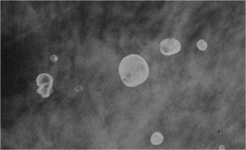
- Oil Cyst - An old focus of fat necrosis that appears as a lucent sphere with thick, calcified rim around it.
 |
 |
Background Information - Imaging Modalities
Mammography remains the gold standard for breast cancer screening and diagnosis; however other imaging modalities and techniques are used in special situations (such as dense breast tissue) or as an adjuvant to better characterize lesions and improve diagnostic accuracy. Below is a table summarizing common imaging modalities.
 |
|
Background Information - Breast Imaging Reporting and Data System (BI-RADS)
BI-RADS is a published standard for breast imaging reporting. It contains definitions of standard reporting terminology, assessment categories, and guidelines for follow-up and monitoring. Final assessment categories range from 0 – 6 as below.
 |
|
Background Information - Types Of Core Biopsies
- Core Needle Biopsy - Can be performed under local anesthesia guided by x-ray, ultrasound, or palpation. Usually multiple passes are made, resulting in as many cores.
- Stereotactic Biopsy - A biopsy that uses imaging in at least two planes to localize a target. Stereotactic guidance can be used for core biopsies or vacuum-assisted biopsies.
- Vacuum-assisted Core Biopsy - A hollow probe is directed through a small cut in the breast under the guidance of x-rays, ultrasound, or MRI. At the target, a cylinder of tissue is pulled by vaccuum into the side of the probe and a rotating knife separates the tissue sample from the rest of the breast. This method allows multiple tissue samples to be collected and yield much more tissue than a standard core biopsy.
Background Information - Specimen Radiography
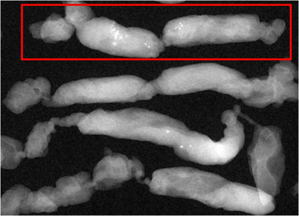
Targeted calcifications are often placed in a special casette. After core biopsy for calcifications, the removed tissue is often imaged to ensure that lesional tissue has been removed and to pinpoint its location in the specimen. The cores with the targeted calcifications are placed in a colored cassette to make correlation as easy as possible. This cassette should be given special attention.
 |
|
Targeted calcifications are often placed in a special casette.