Mucocoele-like Lesion
Contents
Introduction
|
Definition: Mucocoele-like lesions can produce calcifications or a mass evident by radiological imaging or physical examination. The glands associated with mucocoele-like lesions usually possess atypical epithelium. The degree of the atypia must be specified. The presence of atypia in the setting of a mucocoele-like lesion has the same implications that it does in other settings. Clinical Significance: Mucocoele-like lesion is a diagnosis applied to members of a group of lesions characterized by the accumulation of epithelial mucin. In the seminal description, the term referred to collections of mucin within the stroma associated with mucin-filled, distended glands. Latter writings have used the term to refer to a collection of cystic glands filled with mucin without stromal mucin extravasation, but many pathologists still require the presence of stromal mucin pools to render the diagnosis of mucocoele-like lesion. Gross Findings: When visible, mucocoele-like lesions appear as clustered cysts containing translucent, grey, glistening, viscous material. Microscopic Findings: Accumulation of mucin, often accompanied by coarse calcifications, represents the hallmark of mucocoele-like lesions. The mucin collects within ducts and acini and often extravasates into the stroma. Differential Diagnosis: Invasive mucinous carcinoma (clusters of atypical ductal cells in the extravasated mucin pools) Discussion: Because the term mucocoele-like lesion represents only a category of lesions, one must further characterize specific examples by specifying the nature of the epithelium associated with the mucinous material. |
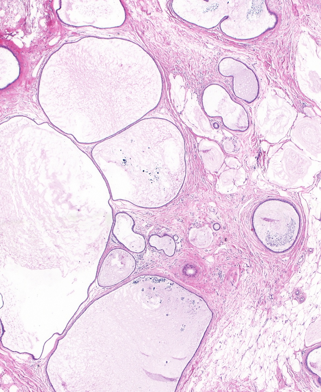 |
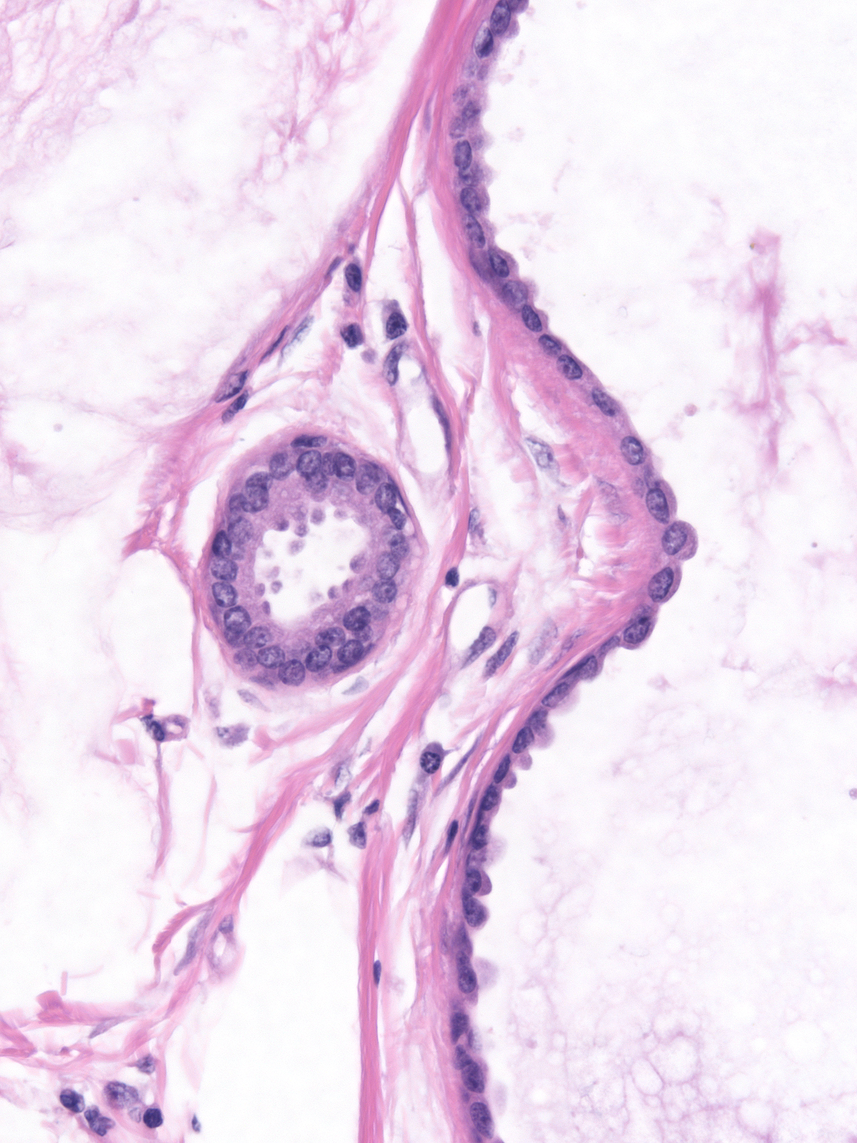
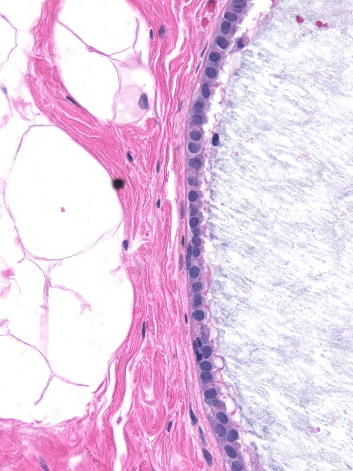
Uncommonly encountered mucocoele-like lesions contain bland flat epithelium or hyperplastic ductal cells.
 |
 |
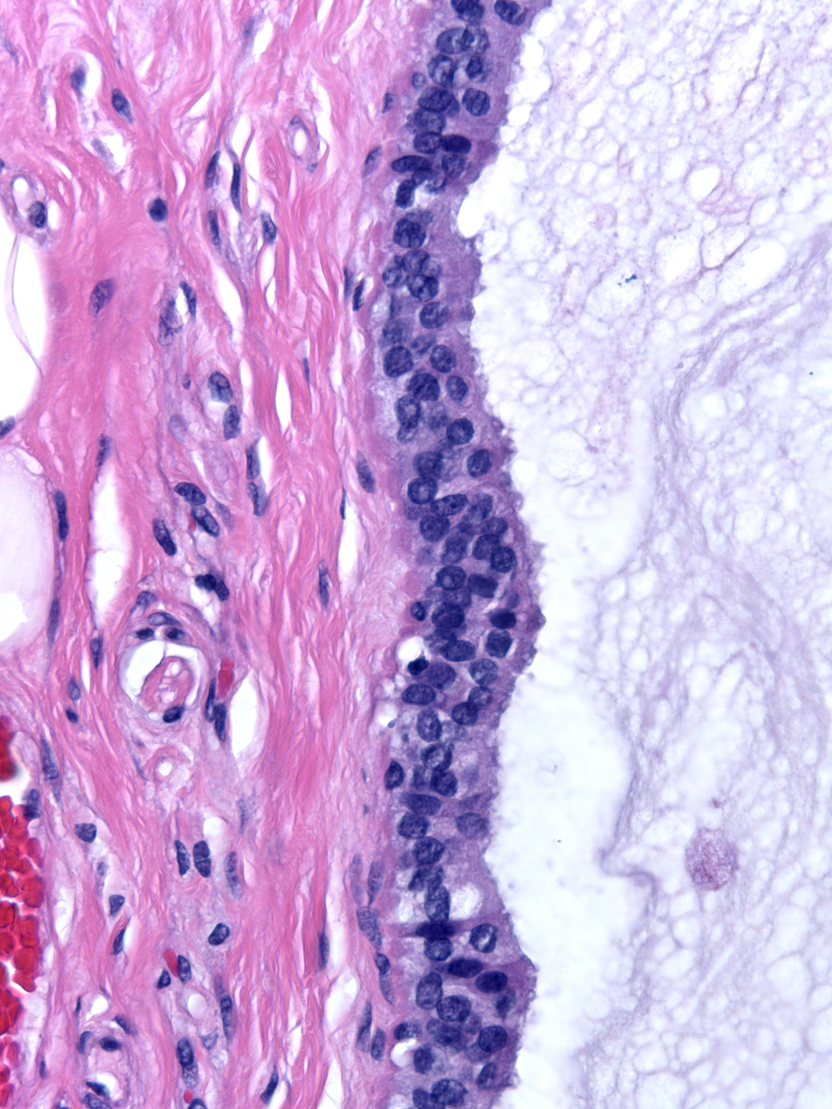
The epithelial cells lining the mucin-containing glands in most mucocoele-like lesions appear atypical. The degree of atypia ranges from low-grade to high-grade. The diagnosis of epithelial atypia in mucocoele-like lesions follows the same guidelines as it does in other settings.
 |
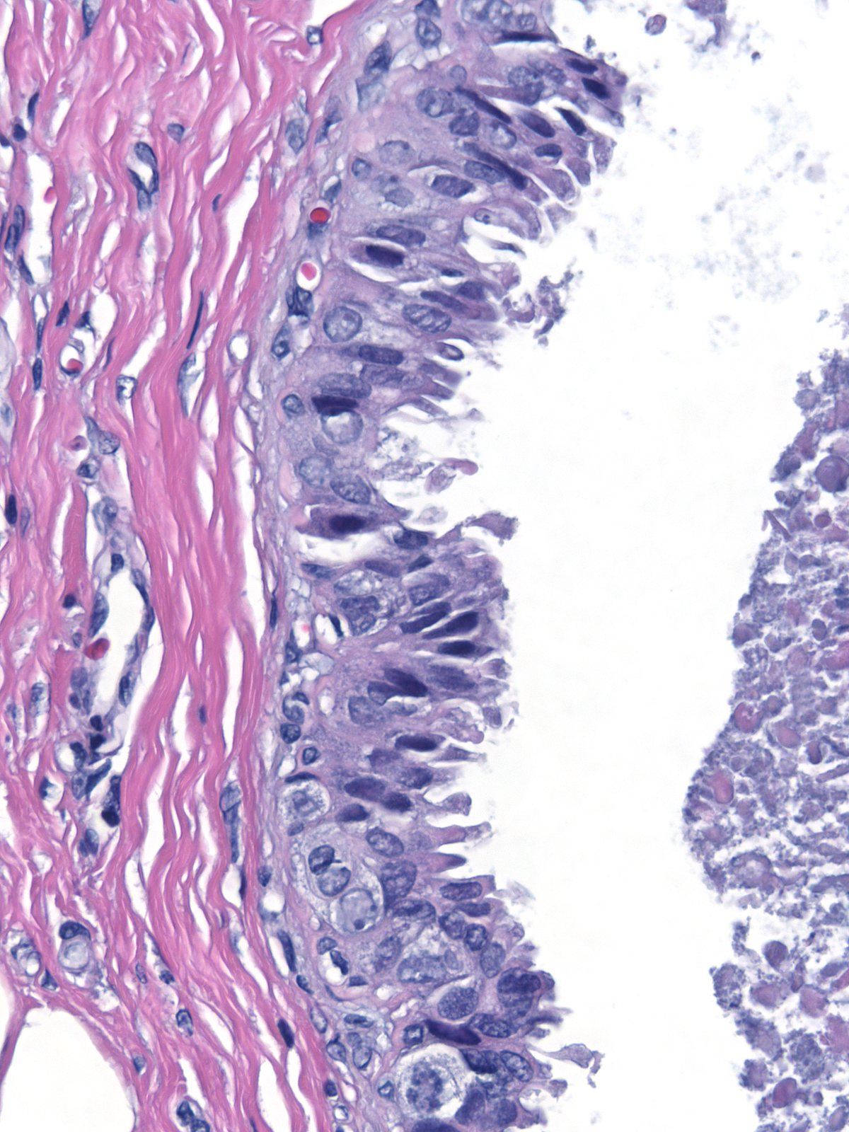 |
The pattern of growth of the epithelial cells duplicates the spectrum seen in the low‑grade pathway. Flat proliferations showing low‑grade atypia merit the diagnosis of flat atypia, whereas stratified, architecturally complex proliferations of similar cells represent atypical ductal hyperplasia or ductal carcinoma in‑situ.
 |
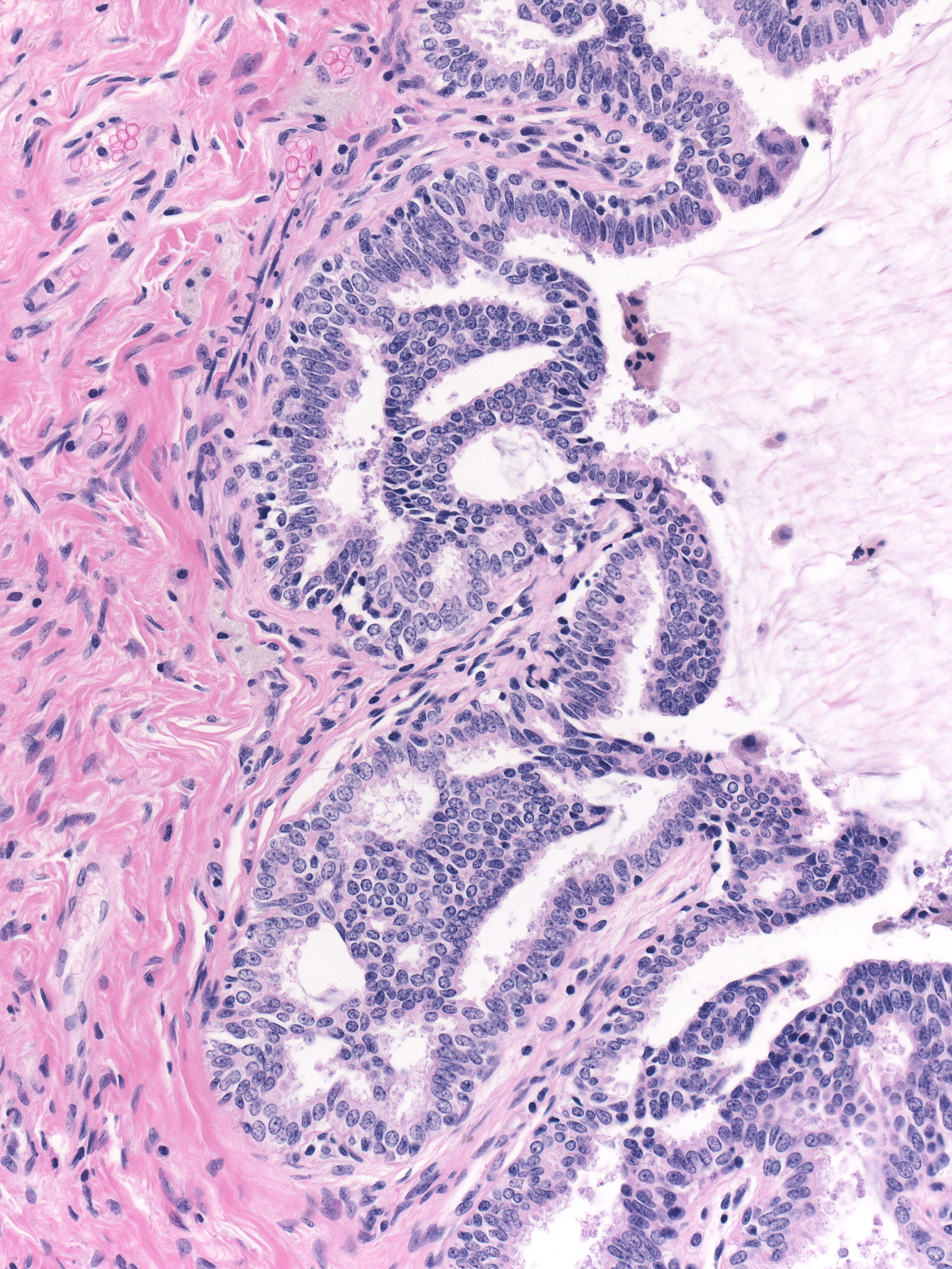 |
| The epithelial proliferation usually has a micropapillary configuration. |  |
| Calcifications often precipitate within the accumulated mucin. They may appear large and pleomorphic on the mammogram. |  |
| When mucin accumulates within glands, it usually collects within the lumen, but it can also dissect between the luminal and myoepithelial cells and between the myoepithelial cells and the basement membrane. |  |
| In cases in which the mucin dissects between the myoepithelial cells and the basement membrane, the appearance superficially resembles the look of extravasated mucin. |  |
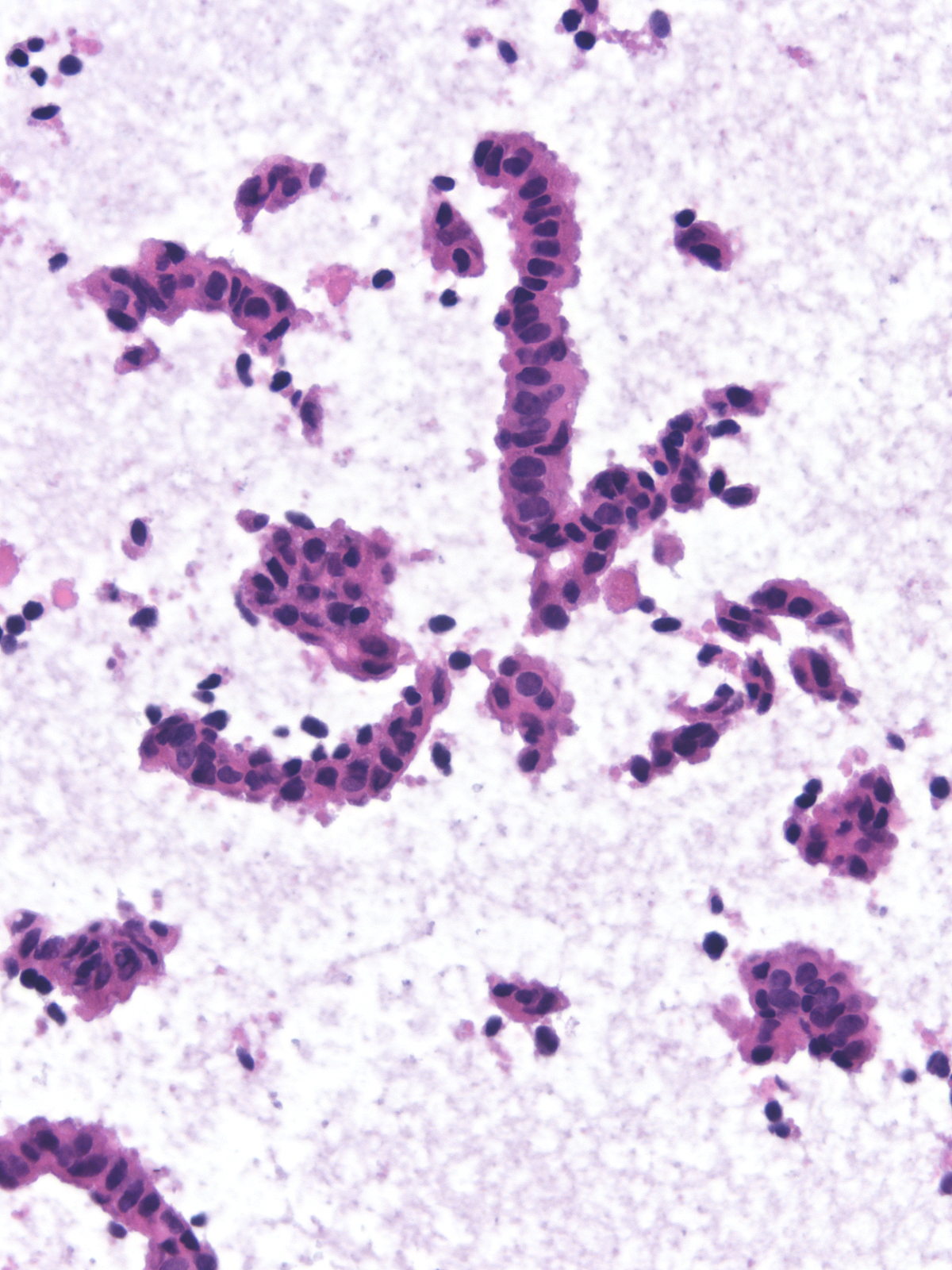
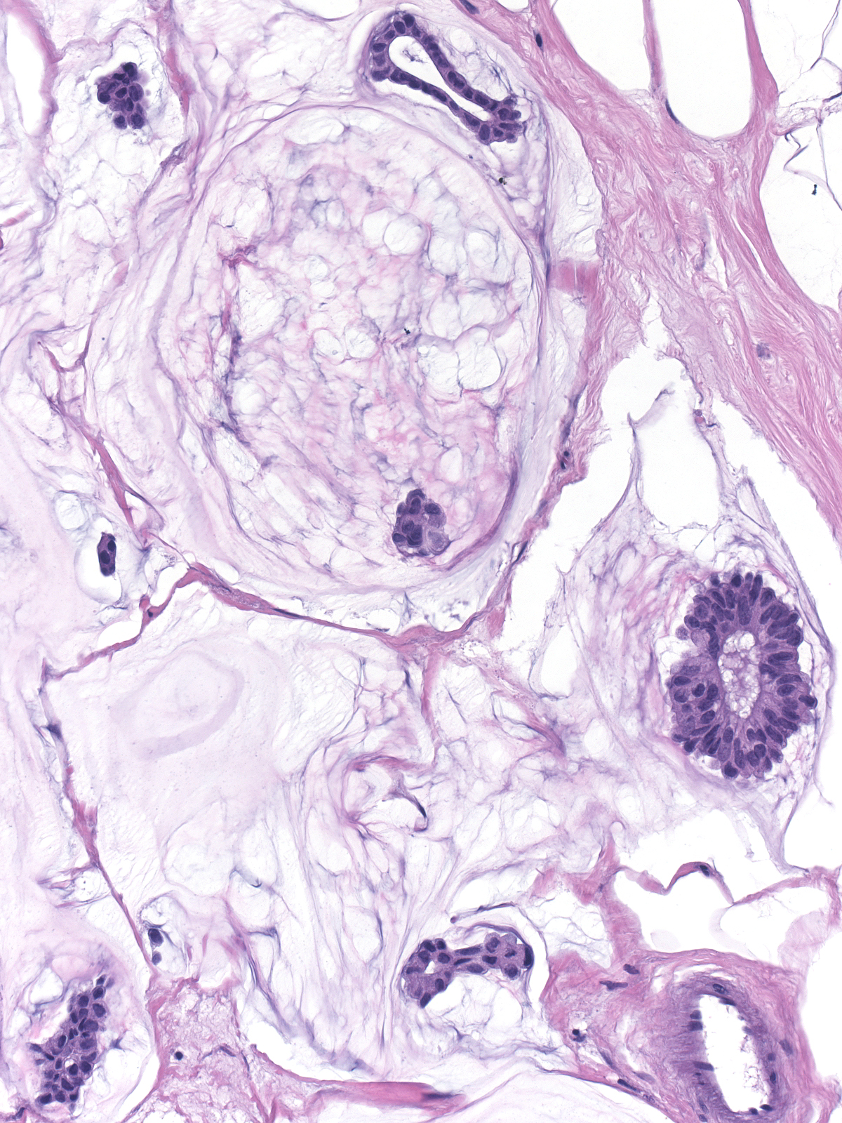
Rough handling during a biopsy procedure or in processing can tear the epithelium from the wall of the affected duct and deposit it within the mucin. The left image illustrates dislodged epithelium within mucin‑filled ducts, whereas the right image shows strips of epithelium in extravasated mucin.
 |
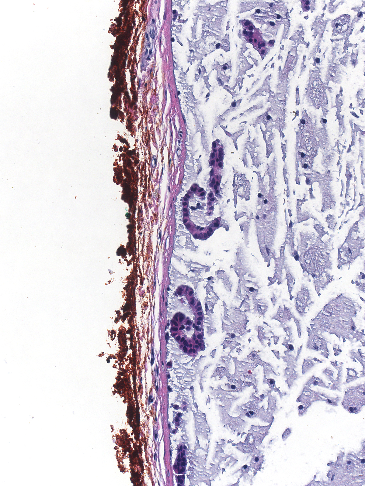 |
The presence of epithelial clusters within mucin can cause one to consider the diagnosis of invasive mucinous carcinoma, but the disrupted single cells, loose shaggy clusters, and linear strands do not look like the intact clusters of invasive mucinous carcinoma. Moreover, when the dislodged epithelium lies within a duct, the shape of the mucin collection and its well defined border identify the location of the mucin as intraductal rather than extravasated.
 |
 |