Tubular Carcinoma
Contents
Introduction
|
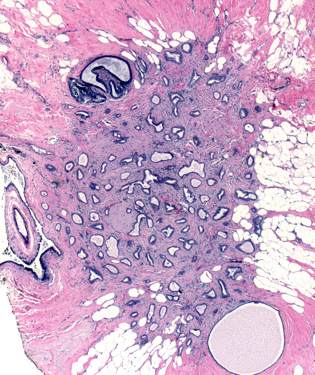
Definition: Tubular carcinoma is a form of well differentiated invasive ductal carcinoma showing low-grade cytology and the formation of simple tubules. Clinical Significance: Tubular carcinoma usually presents as a small mass detected by imaging. These carcinomas metastasize only very rarely; consequently, patients with this form of invasive carcinoma experience an exceptionally favorable prognosis. Gross Findings: Macroscopic examination reveals a small, irregular, hard, white-tan mass. Most measure less than 2.0 cm. Microscopic Findings: The carcinoma consists of angular tubules infiltrating the mammary parenchyma. The cells forming the tubules must form a single layer and exhibit low-grade cytology. Typical tubular carcinomas stain for estrogen receptors. Differential Diagnosis: Sclerosing adenosis (lobulated, zonal organization, intact basement membrane), grade 1 invasive ductal carcinoma NOS (lack of well developed tubules) Discussion: Growth as tubules represents one of the two features required to render the diagnosis of tubular carcinoma. For this purpose, a tubule is defined as a glandular structure with an open lumen formed by a single layer of malignant ductal cells in which every cell touches the lumen. Off-center planes of section may fail to demonstrate the lumen in a few clusters, but the presence of more than a few solid nests, single cells, or linear chains excludes the diagnosis of tubular carcinoma. |
 |
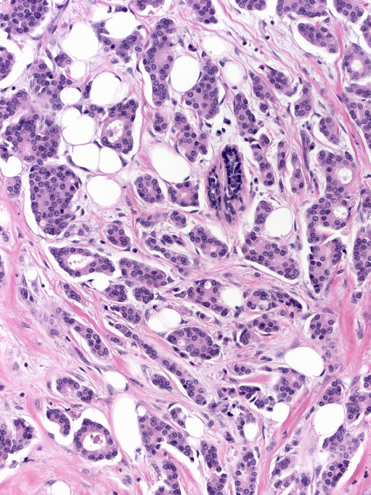
| The cells forming the tubules appear about the size of normal ductal or acinar cells and their nuclei appear about the size of normal nuclei. | 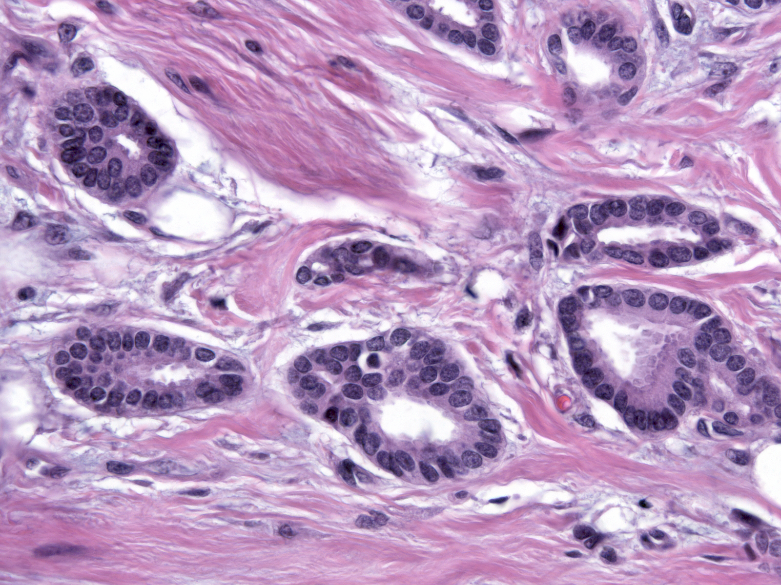 |
| Comparison of the malignant cells with normal ones illustrates the similarities in sizes and shapes. The nuclei contain homogenous dark chromatin and inconspicuous nucleoli. The carcinoma cells possess small amount of eosinophilic cytoplasm. | 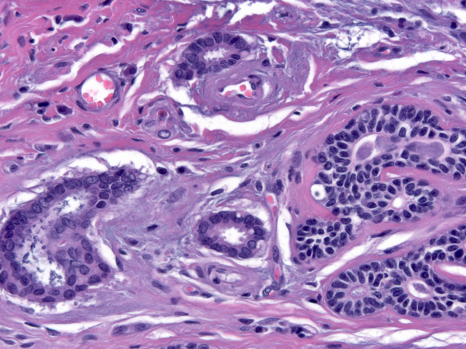 |
| The tubules have irregular shapes; often some have pointed ends. |  |
The stroma typically displays desmoplastic features, but in certain cases it appears essentially unaltered.
 |
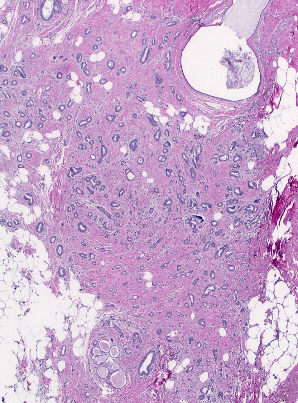 |
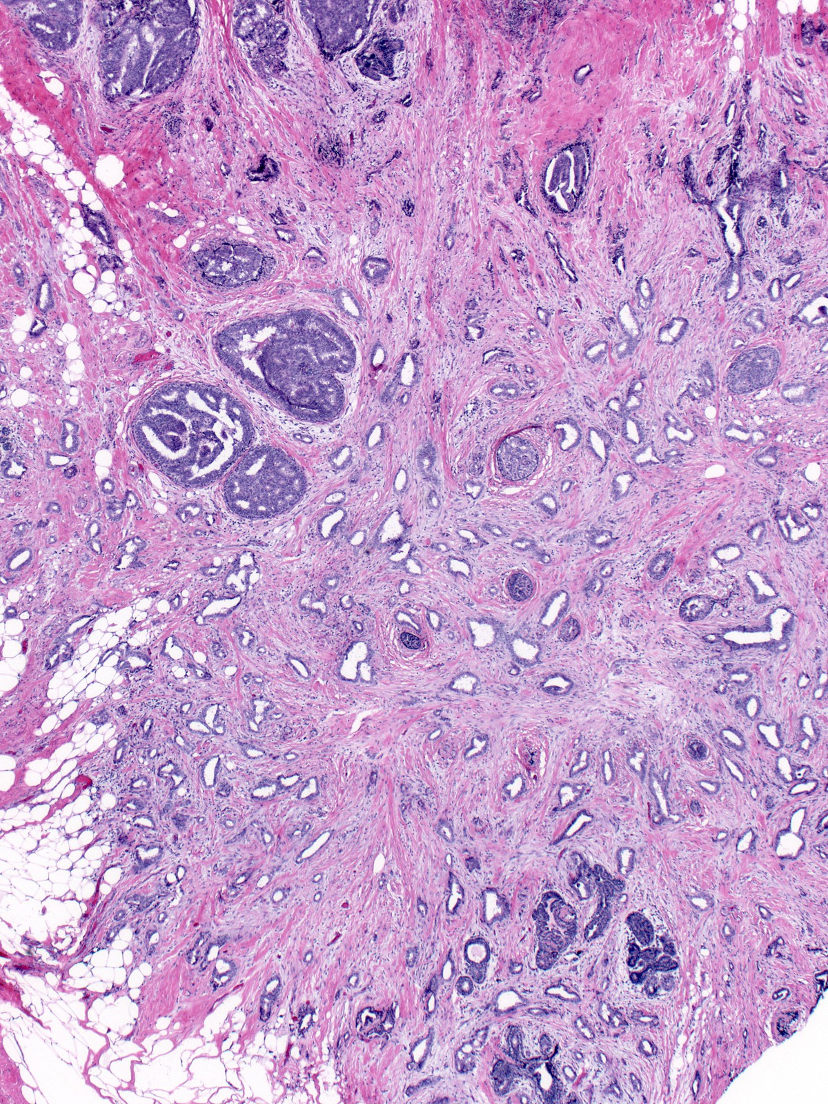
| The arrangement of the tubules does not suggest that they represent distorted pre-existing glandular tissue; instead, they form a space-occupying mass that invades and displaces the underlying tissue. One can detect the phenomenon of invasion most easily at the periphery of the mass. (Invasion of fat occurs in many cases, but one cannot depend on this finding to recognize the presence of invasion.) | 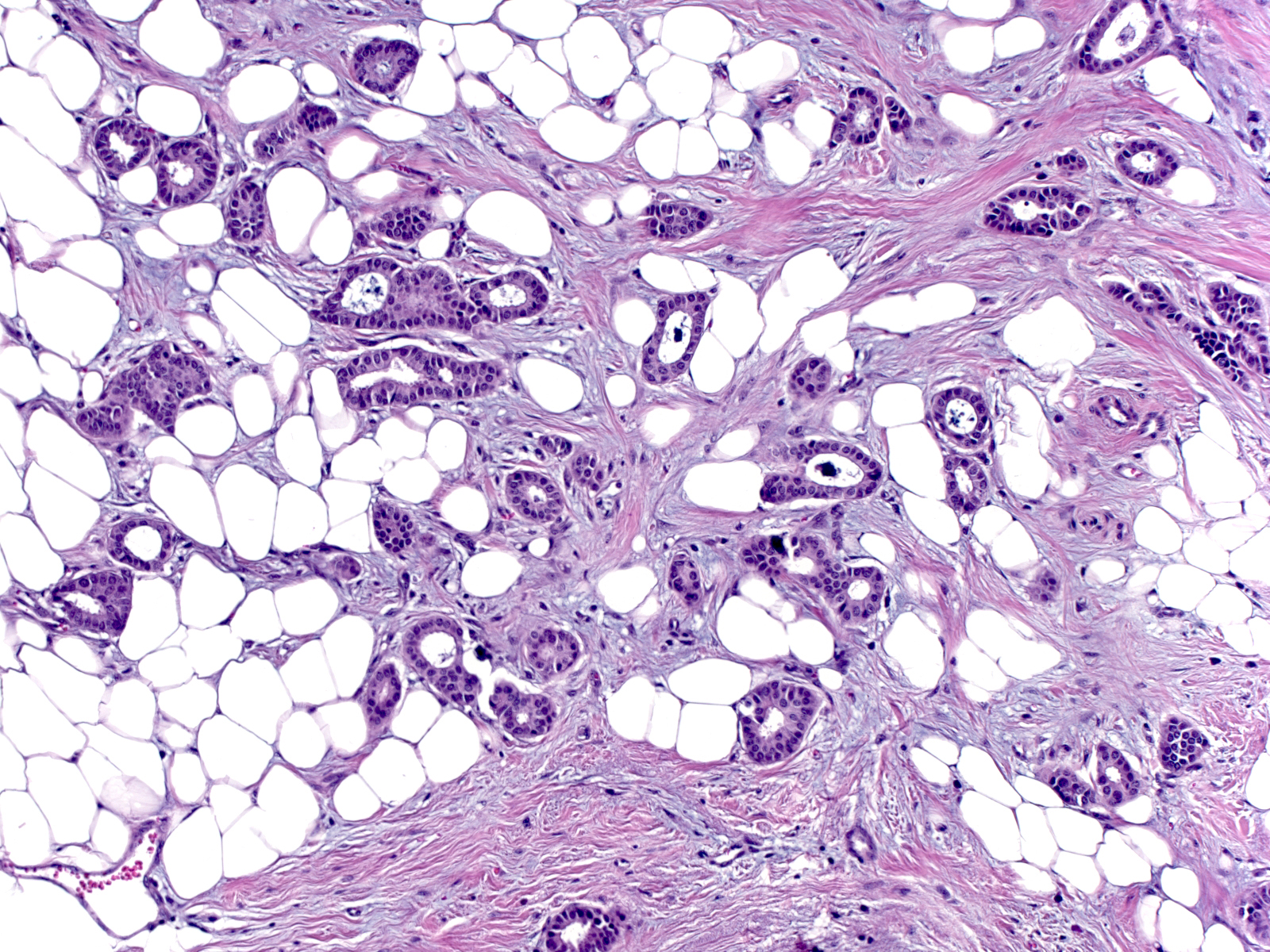 |
| Spread around pre-existing glandular components and disruption of the organization of the collagen bundles represent the most reliable evidence of invasion. | 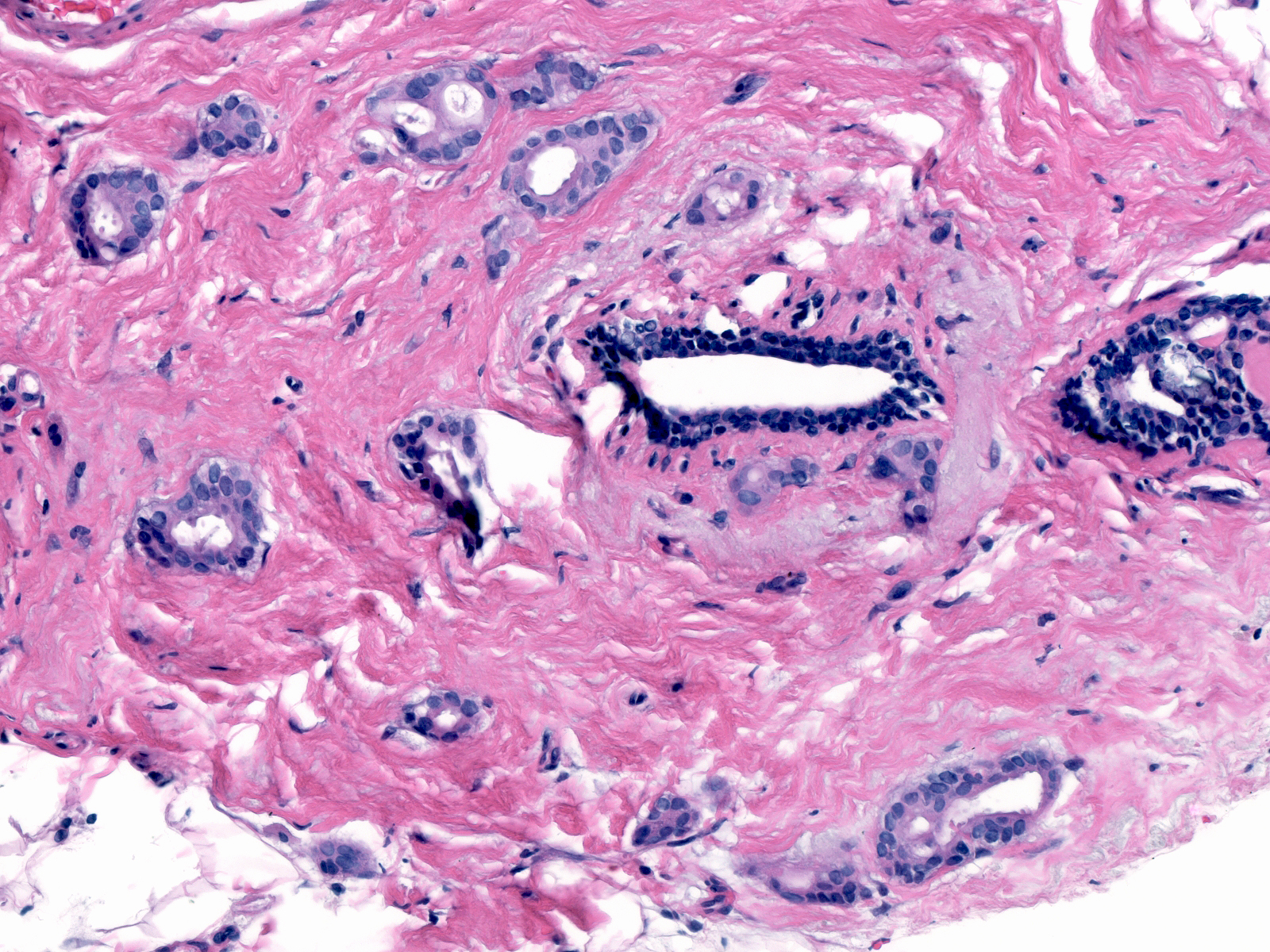 |
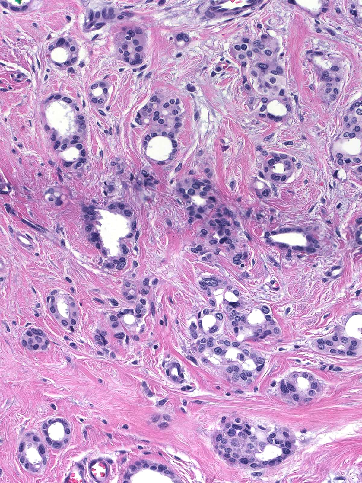
The presence of intermediate‑ or high‑grade atypia excludes the diagnosis of tubular carcinoma, and so does the presence of solid nests or strands of cells.
 |
 |
| Tubular carcinoma frequently co-exists with flat epithelial atypia and lobular neoplasia. Pathologists refer to the constellation composed of tubular carcinoma, lobular neoplasia, and flat atypia as the Rosen triad. (Tubular carcinoma adjacent to FEA ) | 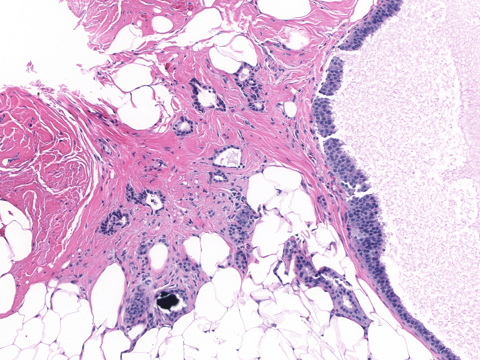 |