General
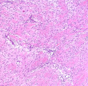
Fibromatosis in the breast demonstrates the same microscopic features of fibromatosis arising at other sites.
- Definition: Fibromatosis is a neoplastic proliferation of bland fibroblasts.
- Clinical Significance: The tumor usually presents as a palpable mass, but small examples may come to attention through radiological imaging. Fibromatosis sometimes arises adjacent to breast implants.
- Gross Findings: Fibromatosis usually forms an ill defined white mass, which may have either a nodular or stellate configuration.
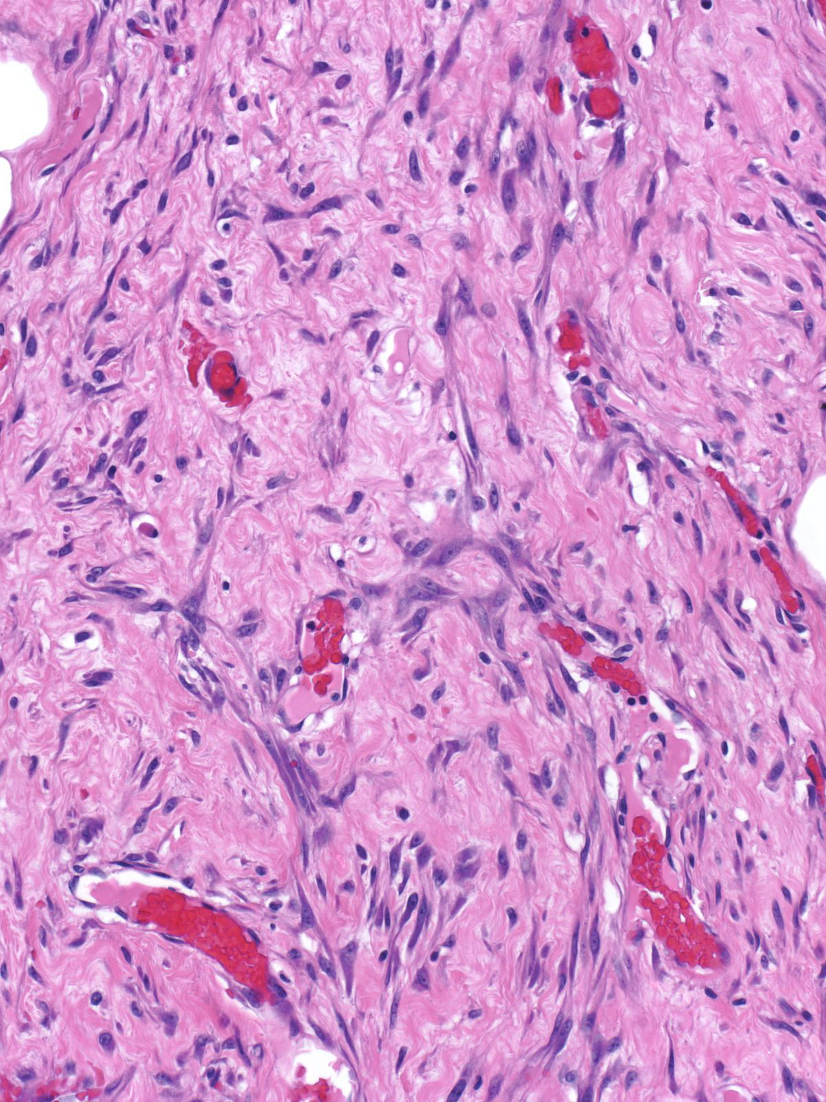
- Microscopic Findings: Mammary fibromatosis demonstrates the microscopical features of fibromatosis arising at other sites. Bland fibroblasts form broad sheets or interlacing fascicles in a collagenous background. The mass contains evenly distributed small blood vessels. Small collections of lymphocytes often dot the periphery of the mass.
- Differential Diagnosis: Spindle cell carcinoma (atypia of neoplastic cells), phyllodes tumor (glandular tissue within the mass)
- Discussion: Proliferation of bland spindle cells and deposition of collagen constitute the fundamental features of fibromatosis. The neoplastic cells form broad sheets or interlacing fascicles, which sometimes create a herring bone pattern.
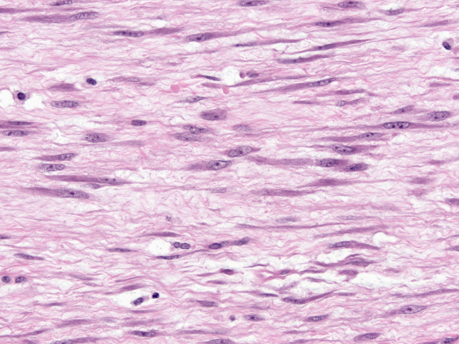
| The cells possess slightly enlarged nuclei, granular chromatin, small nucleoli, and eosinophilic cytoplasm. Mitotic figures are rare. |
 |
|
|
| Bundles of collagen sit among the fibroblasts. Collagen deposition may appear more pronounced at the periphery or in the center. The collagen bundles may appear sparse in certain examples and keloidal in others. In uncommon examples, myxoid material rather than collagen makes up the extracellular matrix. |
 |
|
|
Evenly distributed small blood vessels traverse the mass.
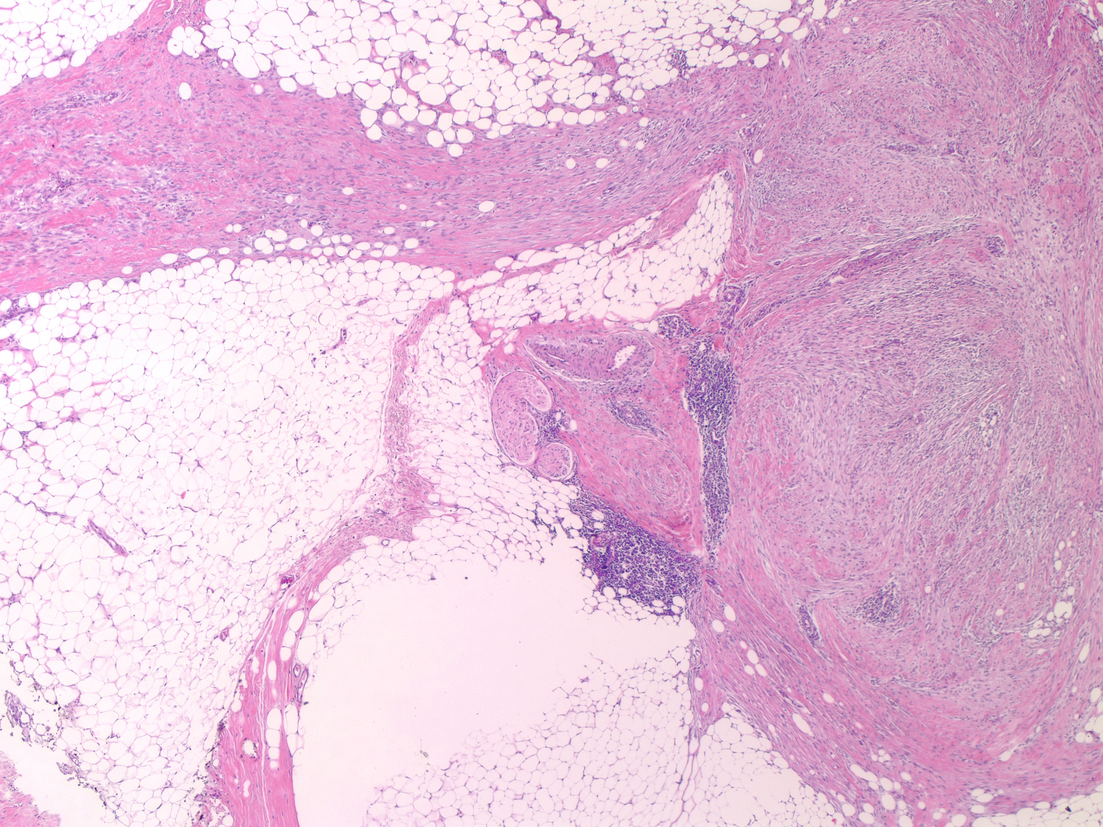
| The fascicles of neoplastic cells extend into the surrounding parenchyma. They may entrap ducts and lobules at the periphery, but one does not find glandular elements deep within the tumor. Small collections of lymphocytes often sit at the periphery of fibromatosis, a useful hint of the diagnosis. |
 |
|
|
| Like cases of extramammary fibromatosis, those arising in the breast often demonstrate nuclear staining for ß-catenin. Demonstration of nuclear ß-catenin reactivity does not establish the diagnosis of mammary fibromatosis, for other spindle cells lesions (such as phyllodes tumor) can demonstrate this finding. |
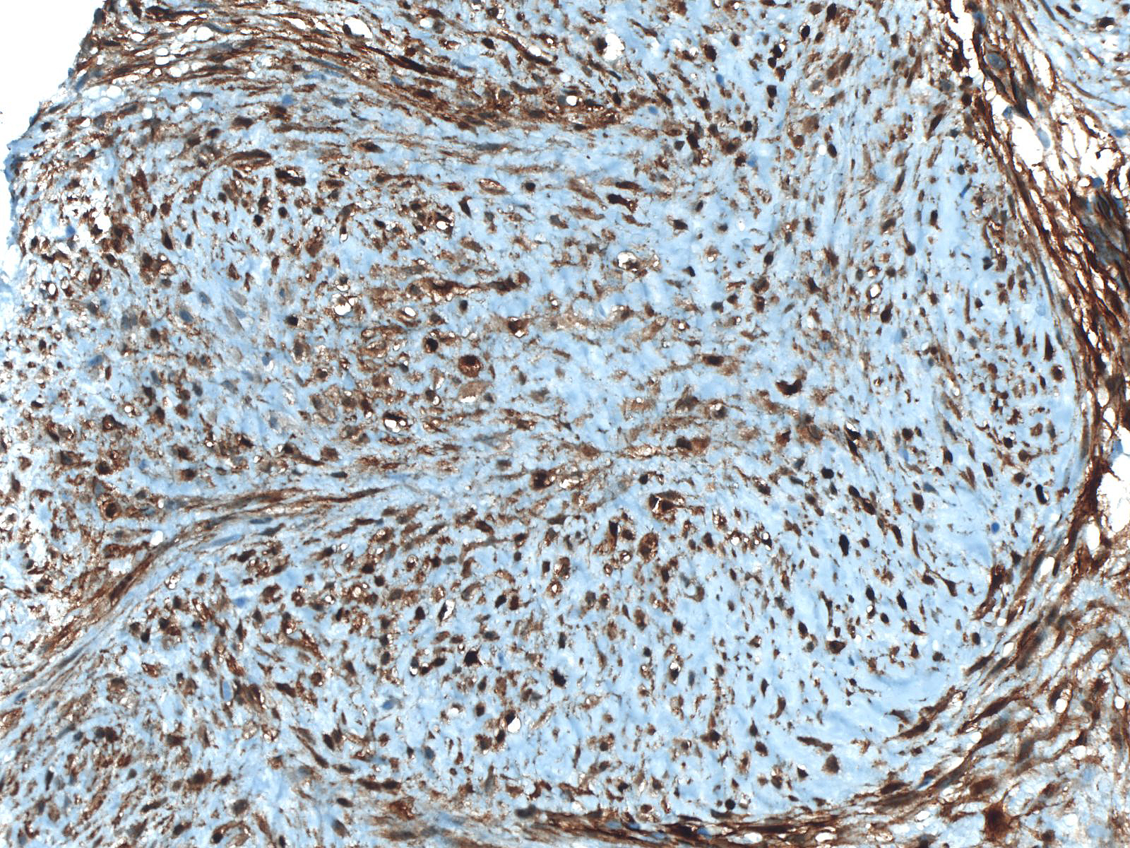 |
|
|
Breast Pathology


