Ductal Carcinoma In Situ
Introduction
|
Definition: Ductal carcinoma in-situ (DCIS) is a neoplastic proliferation of luminal type ductal epithelial cells. Clinical Significance: Ductal carcinoma in-situ comes to clinical attention because of the presence of calcifications on a mammogram or linear enhancement on MRI. In the uncommon situation in which it produces a pronounced stromal reaction, it can present as a mass. DCIS represents the precursor to invasive ductal carcinoma and usually leads to treatment of the entire involved breast (mastectomy or excision and irradiation). Oncologists usually also recommend hormonal therapy to reduce the risk of developing carcinoma in the contralateral breast. Gross Findings: Low-grade DCIS does not usually exhibit macroscopic findings. Transected ducts of high-grade DCIS may exude yellow-white, granular or pasty material (comedonecrosis), corresponding to necrotic carcinoma cells in the duct lumen. Calcifications associated with DCIS may impart a granular texture. Microscopic Findings: Expansive proliferation of polarized, atypical, slightly dishesive epithelial cells characterizes DCIS. Differential Diagnosis: Usual ductal hyperplasia (highly cohesive, nonpolarized cells), atypical ductal hyperplasia (typically low-grade, less expansive), lobular carcinoma in-situ (dishesive, nonpolarized cells) Discussion: Although the cellular features of ductal carcinoma in-situ vary from case to case and can differ somewhat within cases, four alterations represent consistent findings: cellular polarization, cellular enlargement, cytological atypia, and architectural atypia. |
 |
Cellular Polarization
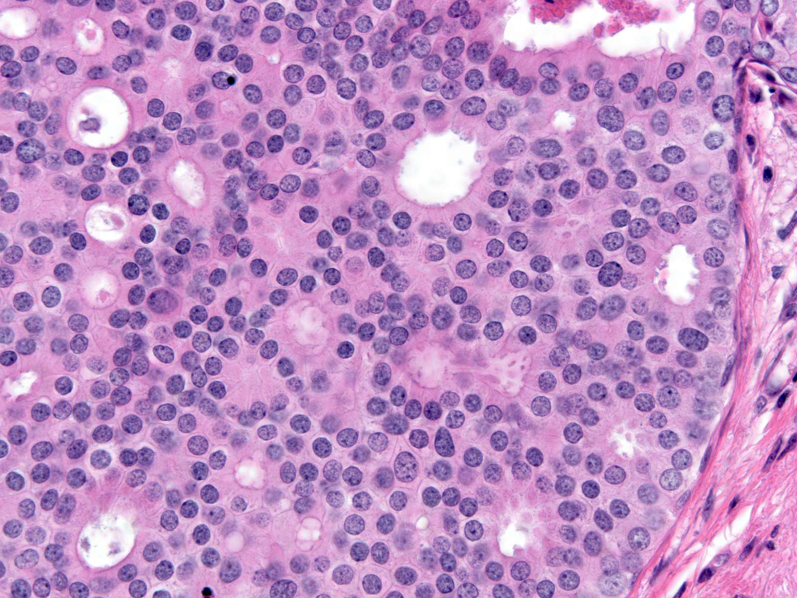
The cells of DCIS display the phenomenon of cellular polarization, a fundamental characteristic of normal epithelial cells. We recognize polarization by the location of the nucleus at one end of the cell (referred to as the basal pole) and the accumulation of cytoplasm at the opposite end (referred to as the apical cytoplasmic compartment). Normal ductal epithelial cells rely on signals associated with the basement membrane to establish the orientation needed to achieve polarity. The ability of ductal carcinoma cells to establish polarity far from the basement membrane differentiates these malignant cells from normal mammary cells and from neoplastic lobular cells.
 |
 |
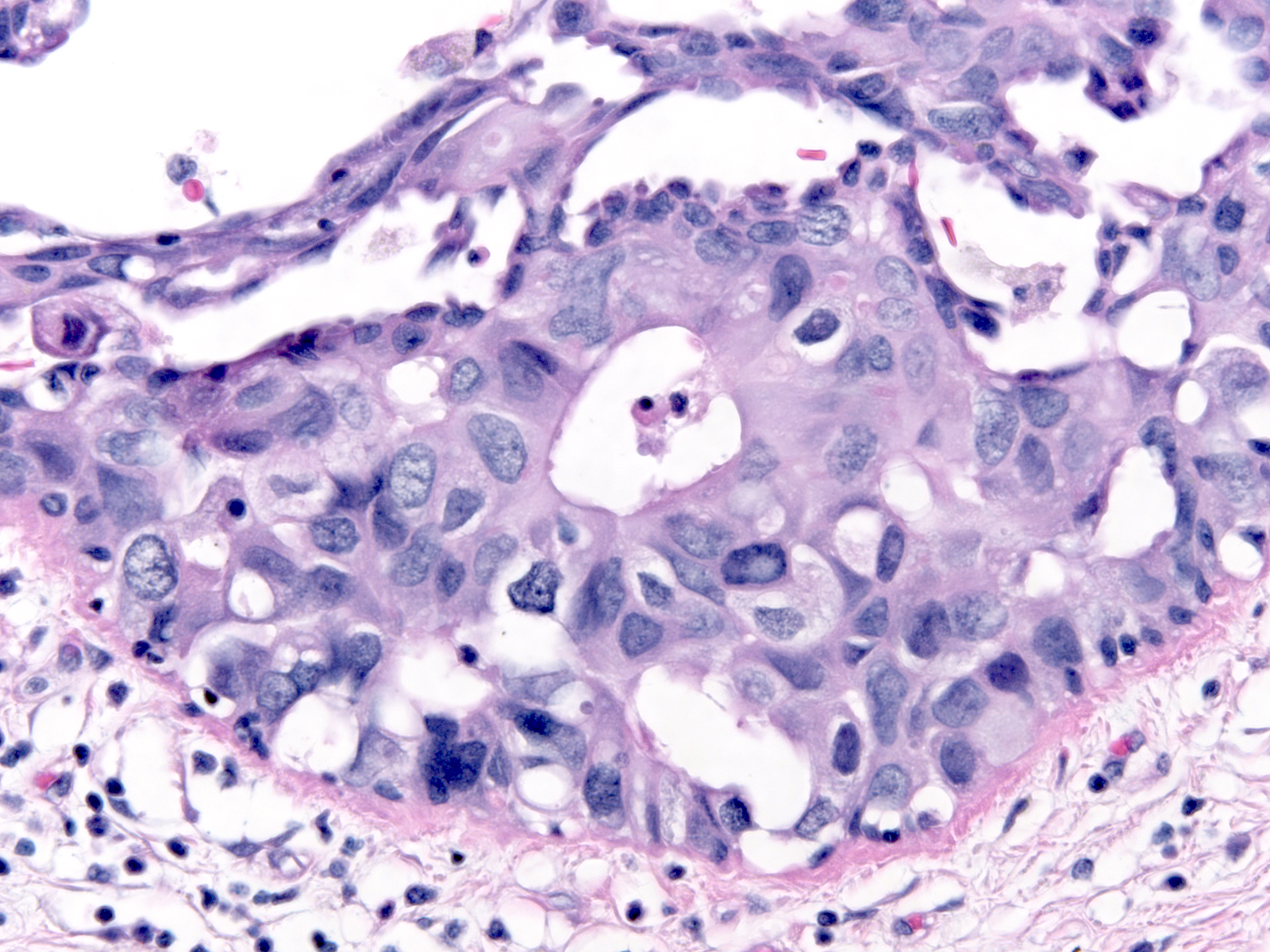
Polarized ductal cells typically display a columnar shape, an eccentric nucleus, and an apical cytoplasmic compartment. Polarization is usually obvious in the cells of low-grade DCIS (above left) and in some cases of intermediate-grade DCIS (above right); however, it may be difficult to appreciate the polarized state in other cases of intermediate-grade DCIS (below left) or in high-grade DCIS (below right). The diagnosis of these later examples of DCIS hinges on recognizing the cells' large size and pronounced cytologic atypia.
 |
 |
Cellular Enlargement
| Besides displaying polarization, the cells of DCIS demonstrate enlargement of the nucleus and increase in the amount of the cytoplasm; thus, the size of the cell increases. Contrasting the malignant cells with nearby benign cells highlights these alterations. In the focus to the right we can easily compare the size of the cells in an entrapped benign gland (center) to the size of the surrounding DCIS cells. | 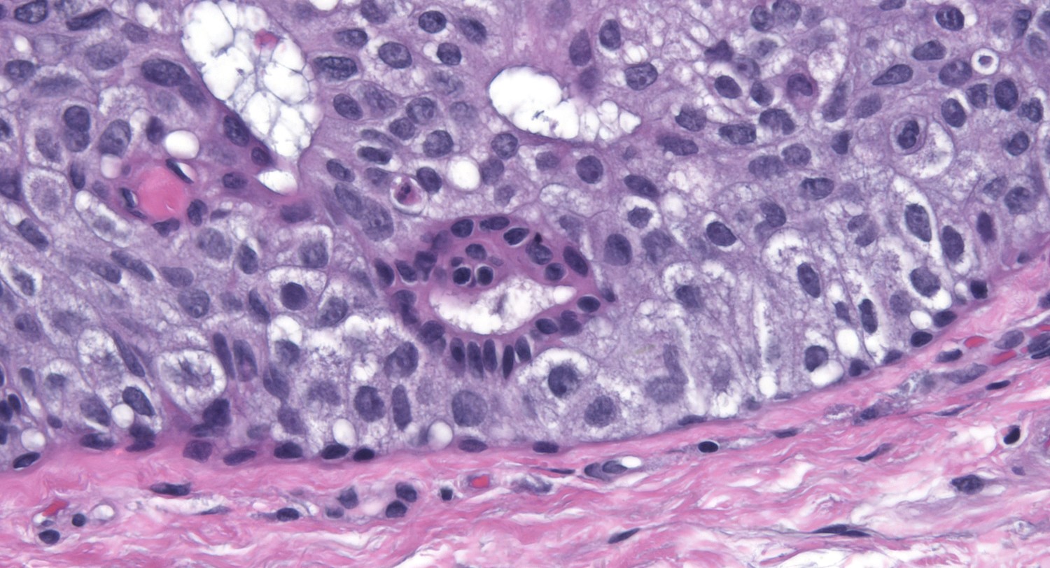 |
Cytologic Atypia
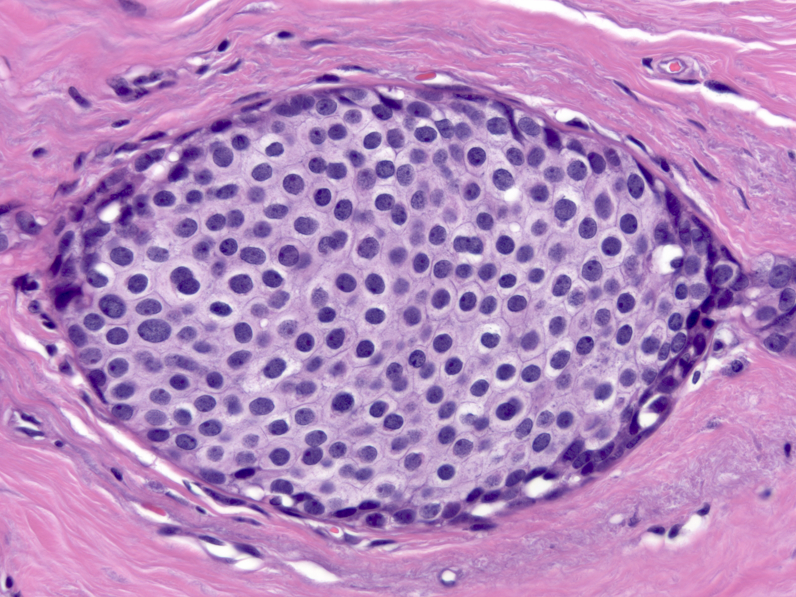
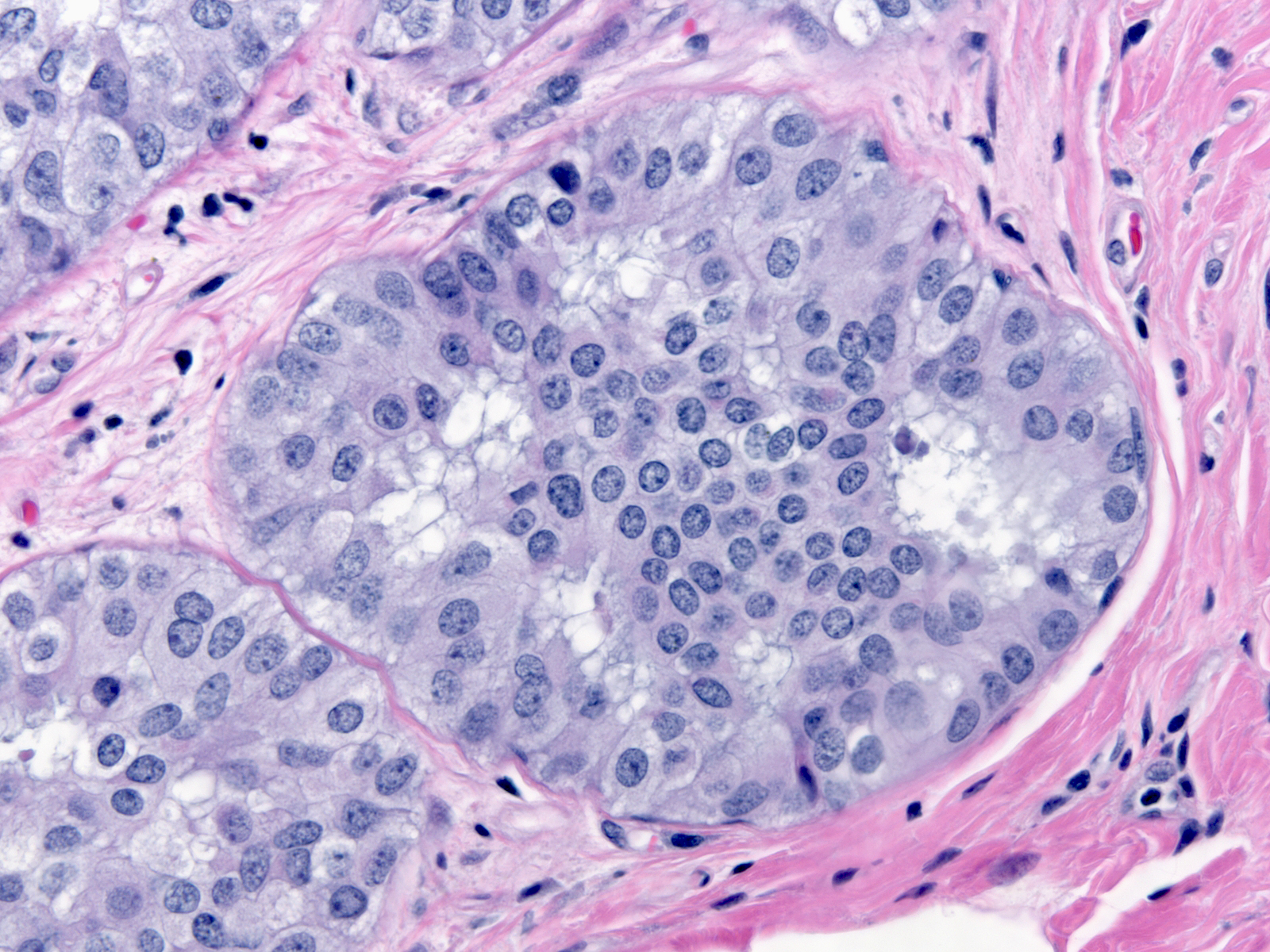
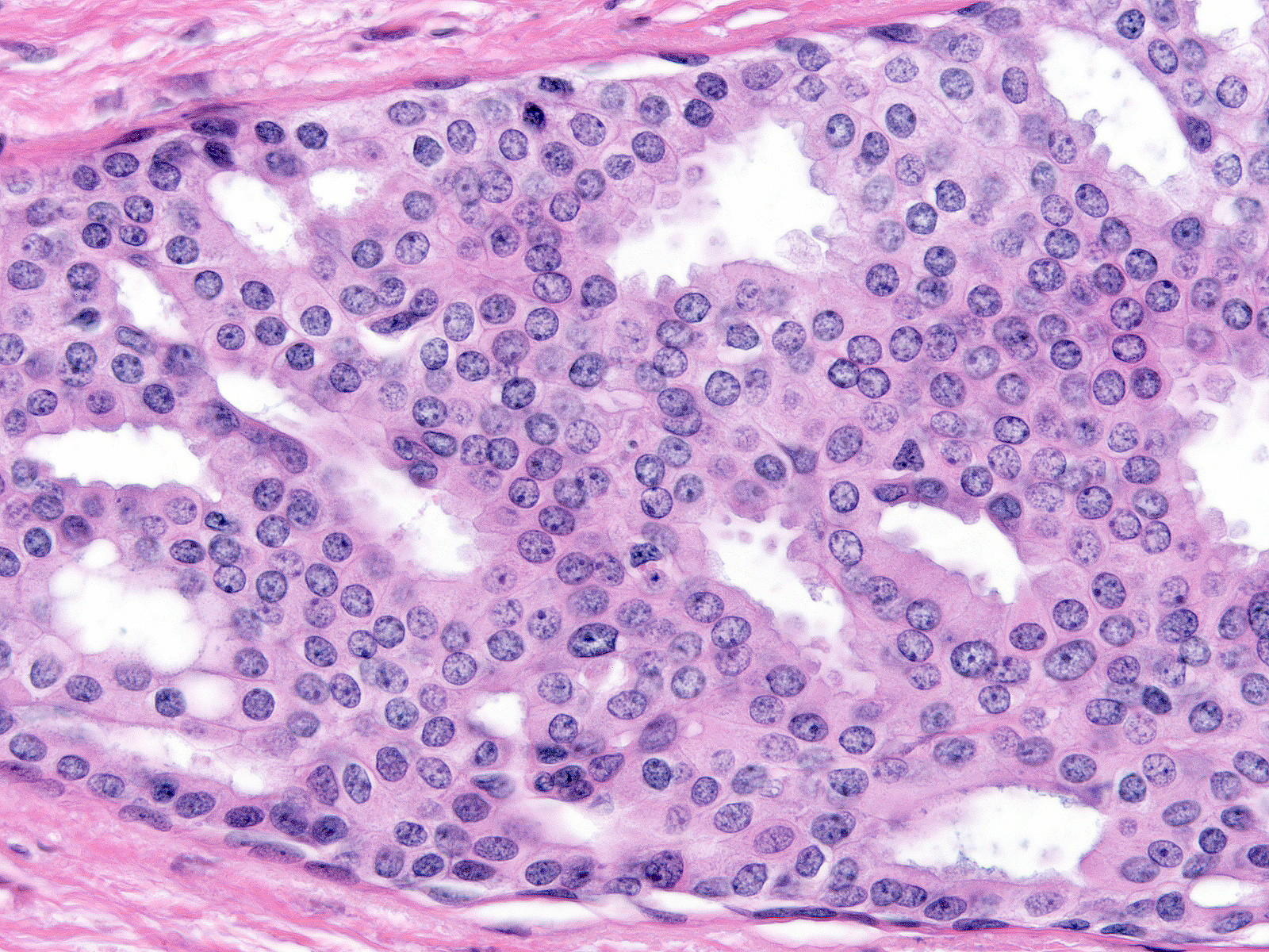
| The cytological atypia that characterizes DCIS varies according to the grade of the carcinoma. Features of low-grade atypia include: uniformity of the nuclei, round or oval nuclear shapes, smooth nuclear contours, and inconspicuous nucleoli. | 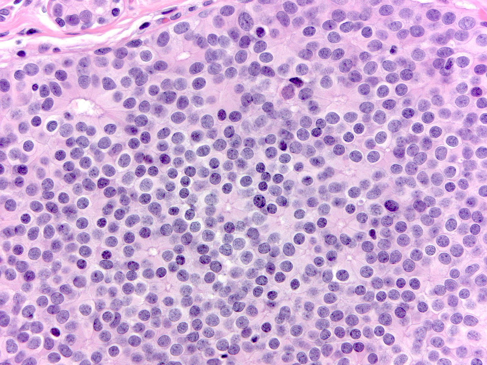 |
The chromatin of low-grade atypia can appear dark and dispersed (below left) or somewhat pale and finely granular (below middle). When it is pale and finely granular, you can usually see small nucleoli (below right).
 |
 |
 |