Myxoid fibroadenoma is a type of fibroepithelial tumor that arises from accumulation of myxoid extracellular matrix produced by fibroblasts of the specialized stroma.
Contents
Introduction
Warning: Display title "<span style="color:#AF0C3E"><b><font size="5"><span style="background-color:; color:">Myxoid fibroadenoma is a type of fibroepithelial tumor that arises from accumulation of myxoid extracellular matrix produced by fibroblasts of the specialized stroma.</span></font></b></span>" overrides earlier display title "Myxoid Fibroadenoma".
|
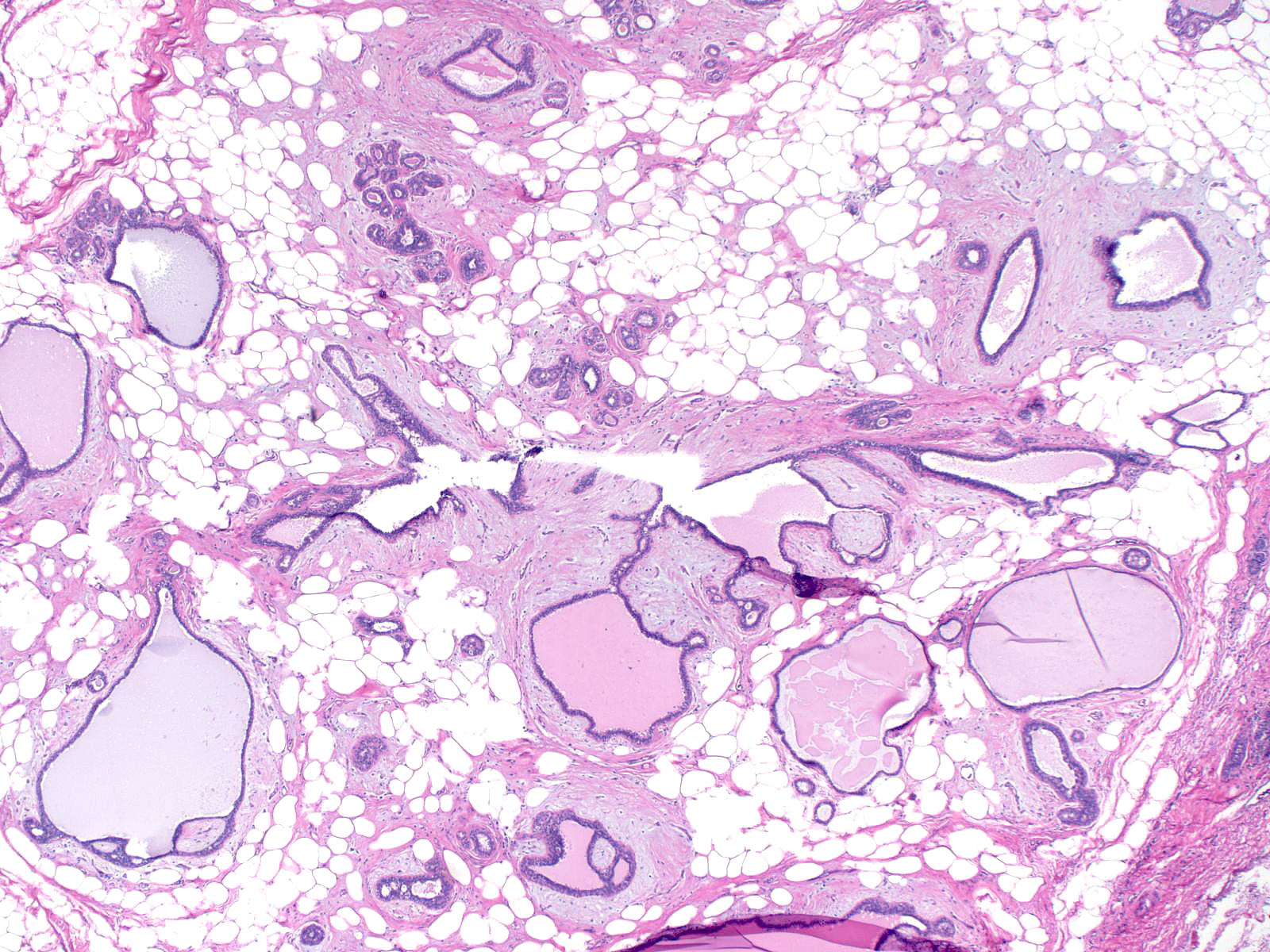
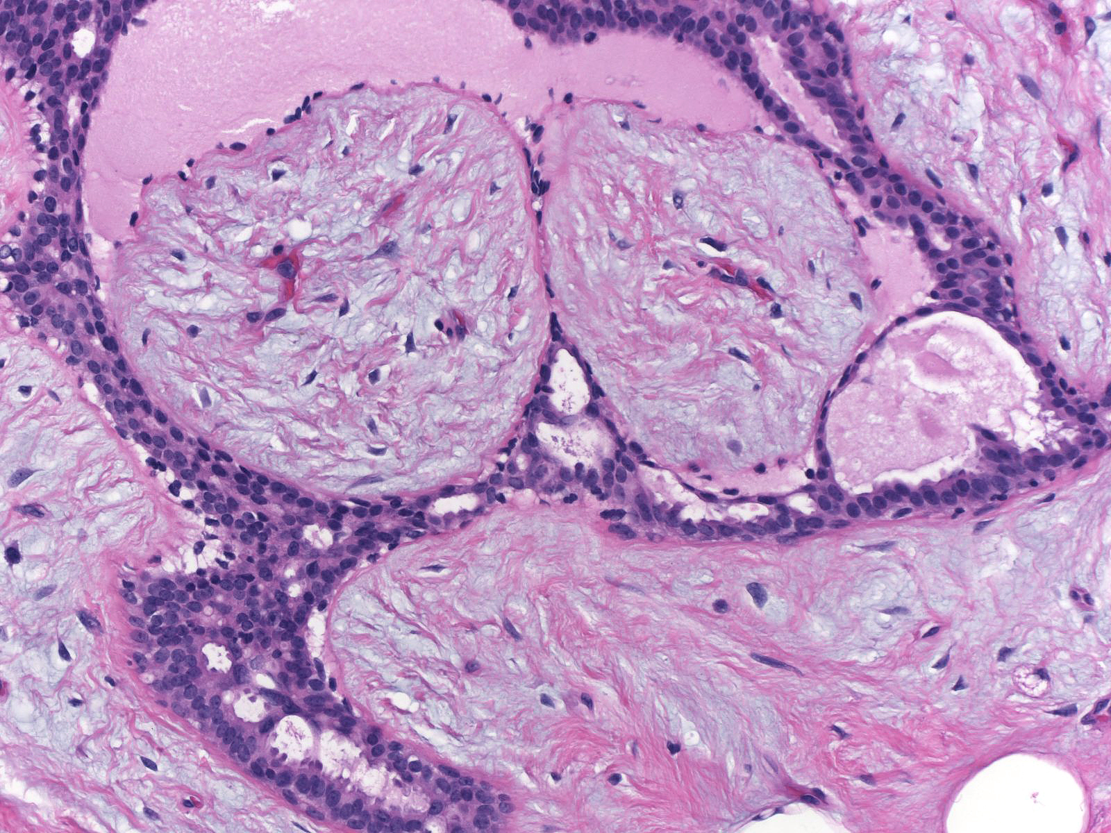
Definition: Myxoid fibroadenomas tend to occur in women beyond the age of thirty years. They sometimes occur as a component of the Carney complex. Clinical Significance: These nodules appear well-defined, grey, and glistening. The tissue often bulges from the cut surface. Gross Findings: Myxoid fibroadenomas consists of abundant blue-grey extracellular matrix surrounding entrapped ducts, ductules, and terminal duct-lobular units. Microscopic Findings: Fibroadenoma with myxoid stroma (proliferation of fibroblasts), myxoid hamartoma (disorganized architecture), myxoid phyllodes tumor (aggressive proliferation of fibroblasts), myxoma (no epithelial component). (A myxoma is a rare lesion that consists of a mass-like collection of stromal mucin devoid of glandular elements.) Differential Diagnosis: Although use of the term, myxoid fibroadenoma, suggests that this lesion represents a variety of fibroadenoma, the pathogeneses of the two entities differ. Discussion: 30-1 Myxoid_FBA_-_intro.jpg |
| Proliferation of fibroblasts gives rise to fibroadenomas, whereas accumulation of myxoid ground substances accounts for the formation of myxoid fibroadenomas. |  |
The extracellular material fills and enlarges the specialized stromal compartment surrounding ducts and lobules. The expansion spreads apart the acini within the lobules.
 |
 |
| Often the extracellular material forms collections that look like nodules protruding into ducts. |  |
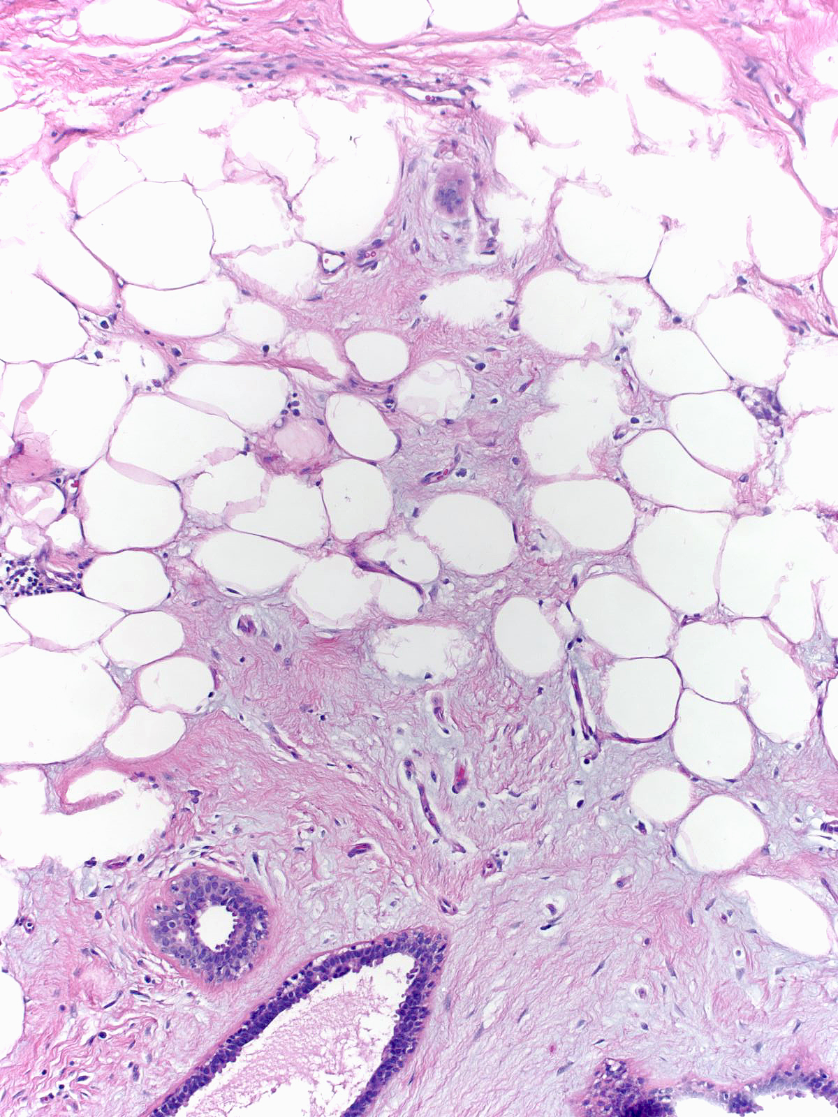
The material may also penetrate into fat and fibrous connective tissue; consequently, adipocytes often become incorporated into the mass and the border of the mass may appear ill defined.
 |
 |
| The glands within the nodule may become atrophic, and the epithelium lining them may appear attenuated. The myoepithelial cells may look especially stretched and the basement membrane often appears thick. With age, the myxoid material undergoes hyalinization and transforms into sparsely cellular collagen. |  |
The presence of abundant myxoid extracellular material does not, itself, establish the diagnosis of myxoid fibroadenoma, for abundant myxoid material can accumulate in typical fibroadenomas, hamartomas, and phyllodes tumors. One differentiates these lesions from myxoid fibroadenomas on the basis of the number and nature of the stromal cells and the presence of their other characteristic findings.
 |
 |
 |