Difference between revisions of "Adenoid Cystic Carcinoma"
| Line 3: | Line 3: | ||
<br> | <br> | ||
== Introduction == | == Introduction == | ||
| − | {{dxintronodisc|Adenoid Cystic Carcinoma|Adenoid cystic carcinoma is a type of invasive ductal carcinoma that features two types of neoplastic cells: luminal and basaloid/myoepithelial.|Adenoid cystic carcinoma usually present as a single, small, palpable mass, which consist predominantly of invasive nests. If present, the noninvasive component represents only a minor component of the carcinoma. This type of carcinoma almost never metastasizes; consequently patients with adenoid cystic carcinoma experience a more favorable prognosis compared to those with conventional invasive ductal carcinomas. Like their salivary gland counterparts, mammary adenoid cystic carcinomas usually contain the MYB-NFIB fusion gene.|The carcinoma usually forms a well defined grey, tan, or pink nodule between one and three centimeters in dimension.|Geographic islands of neoplastic cells infiltrate the mammary parenchyma. Careful evaluation reveals two cell types: luminal epithelial and basaloid/myoepithelial cells. The typical adenoid cystic carcinoma does not stain for estrogen receptors. | + | {{dxintronodisc|Adenoid Cystic Carcinoma|Adenoid cystic carcinoma is a type of invasive ductal carcinoma that features two types of neoplastic cells: luminal and basaloid/myoepithelial.|Adenoid cystic carcinoma usually present as a single, small, palpable mass, which consist predominantly of invasive nests. If present, the noninvasive component represents only a minor component of the carcinoma. This type of carcinoma almost never metastasizes; consequently patients with adenoid cystic carcinoma experience a more favorable prognosis compared to those with conventional invasive ductal carcinomas. Like their salivary gland counterparts, mammary adenoid cystic carcinomas usually contain the MYB-NFIB fusion gene.|The carcinoma usually forms a well defined grey, tan, or pink nodule between one and three centimeters in dimension.|Geographic islands of neoplastic cells infiltrate the mammary parenchyma. Careful evaluation reveals two cell types: luminal epithelial and basaloid/myoepithelial cells. The typical adenoid cystic carcinoma does not stain for estrogen receptors. Cribriform carcinoma (Cribriform carcinoma is a rare form of low‑grade invasive ductal carcinoma in which the neoplastic cells grow in large nests and form glandular spaces identical those found in cribriform ductal carcinoma in-‑situ. The neoplastic cells display the features of luminal cells only and they express estrogen receptors.)|19-1 Low_power-2.jpg|Adenoid cystic carcinoma can have a characteristic low-power appearance; however, the diagnosis depends on recognizing two lines of differentiation of the neoplastic cells.}} |
{{img1|The diagnosis of mammary adenoid cystic carcinoma rests on the presence of a single neoplastic population in which certain carcinoma cells show attributes of luminal cells and others display features of basaloid/myoepithelial cells.|19-2 Invasion.jpg}} | {{img1|The diagnosis of mammary adenoid cystic carcinoma rests on the presence of a single neoplastic population in which certain carcinoma cells show attributes of luminal cells and others display features of basaloid/myoepithelial cells.|19-2 Invasion.jpg}} | ||
Revision as of 08:19, July 17, 2020
Contents
Introduction
|
Definition: Adenoid cystic carcinoma is a type of invasive ductal carcinoma that features two types of neoplastic cells: luminal and basaloid/myoepithelial. Clinical Significance: Adenoid cystic carcinoma usually present as a single, small, palpable mass, which consist predominantly of invasive nests. If present, the noninvasive component represents only a minor component of the carcinoma. This type of carcinoma almost never metastasizes; consequently patients with adenoid cystic carcinoma experience a more favorable prognosis compared to those with conventional invasive ductal carcinomas. Like their salivary gland counterparts, mammary adenoid cystic carcinomas usually contain the MYB-NFIB fusion gene. Gross Findings: The carcinoma usually forms a well defined grey, tan, or pink nodule between one and three centimeters in dimension. Microscopic Findings: Geographic islands of neoplastic cells infiltrate the mammary parenchyma. Careful evaluation reveals two cell types: luminal epithelial and basaloid/myoepithelial cells. The typical adenoid cystic carcinoma does not stain for estrogen receptors. Cribriform carcinoma (Cribriform carcinoma is a rare form of low‑grade invasive ductal carcinoma in which the neoplastic cells grow in large nests and form glandular spaces identical those found in cribriform ductal carcinoma in-‑situ. The neoplastic cells display the features of luminal cells only and they express estrogen receptors.) Differential Diagnosis: 19-1 Low_power-2.jpg |
| The diagnosis of mammary adenoid cystic carcinoma rests on the presence of a single neoplastic population in which certain carcinoma cells show attributes of luminal cells and others display features of basaloid/myoepithelial cells. | 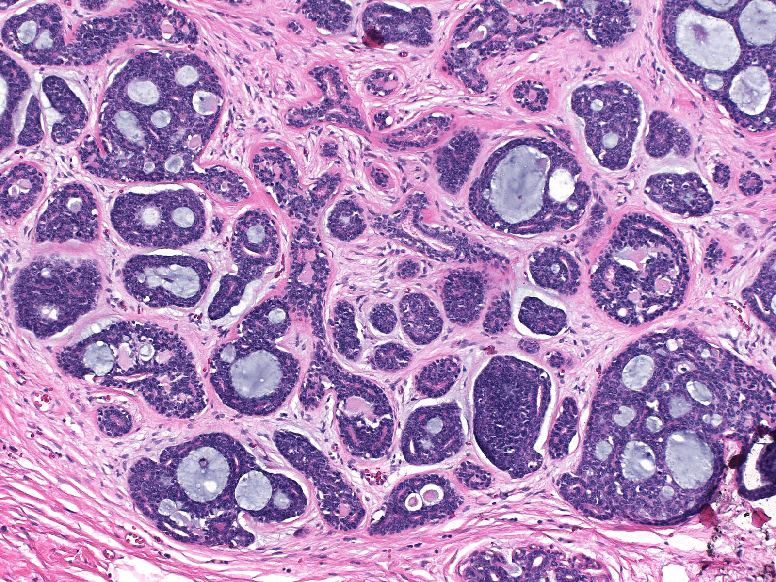 |
| The luminal cells appear polarized and radially arrayed around a lumen. They possess basally situated nuclei containing granular chromatin and small nucleoli and eosinophilic apical cytoplasm. The basaloid/myoepithelial cells do not demonstrate polarization. They have oval spindly nuclei and inconspicuous nucleoli. The nuclei appear circumferentially oriented with respect to spaces formed by the cells. | 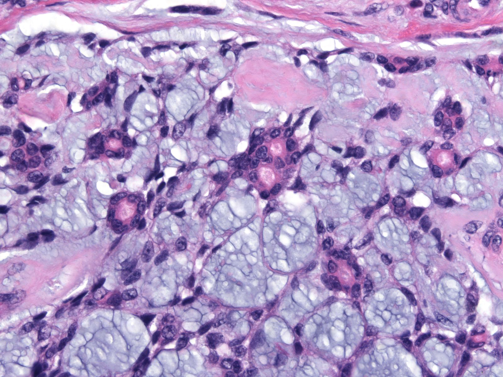 |
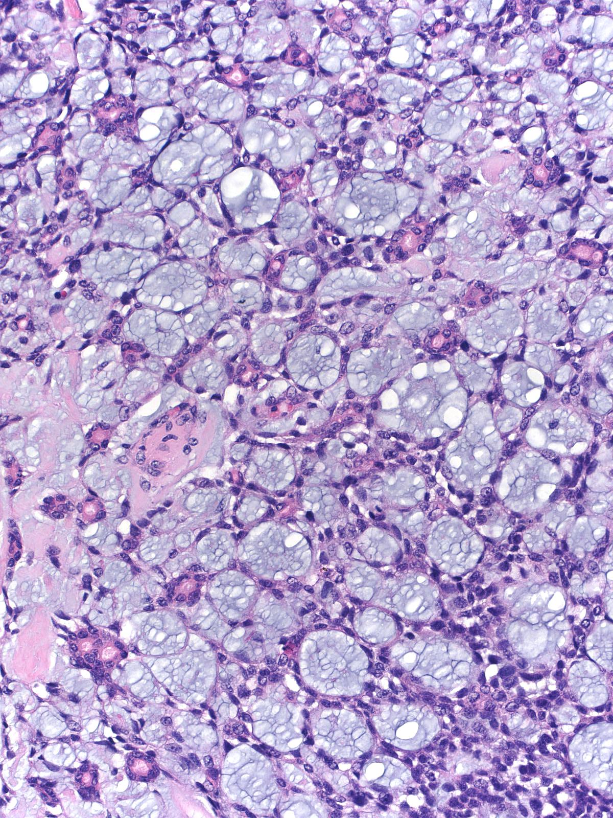
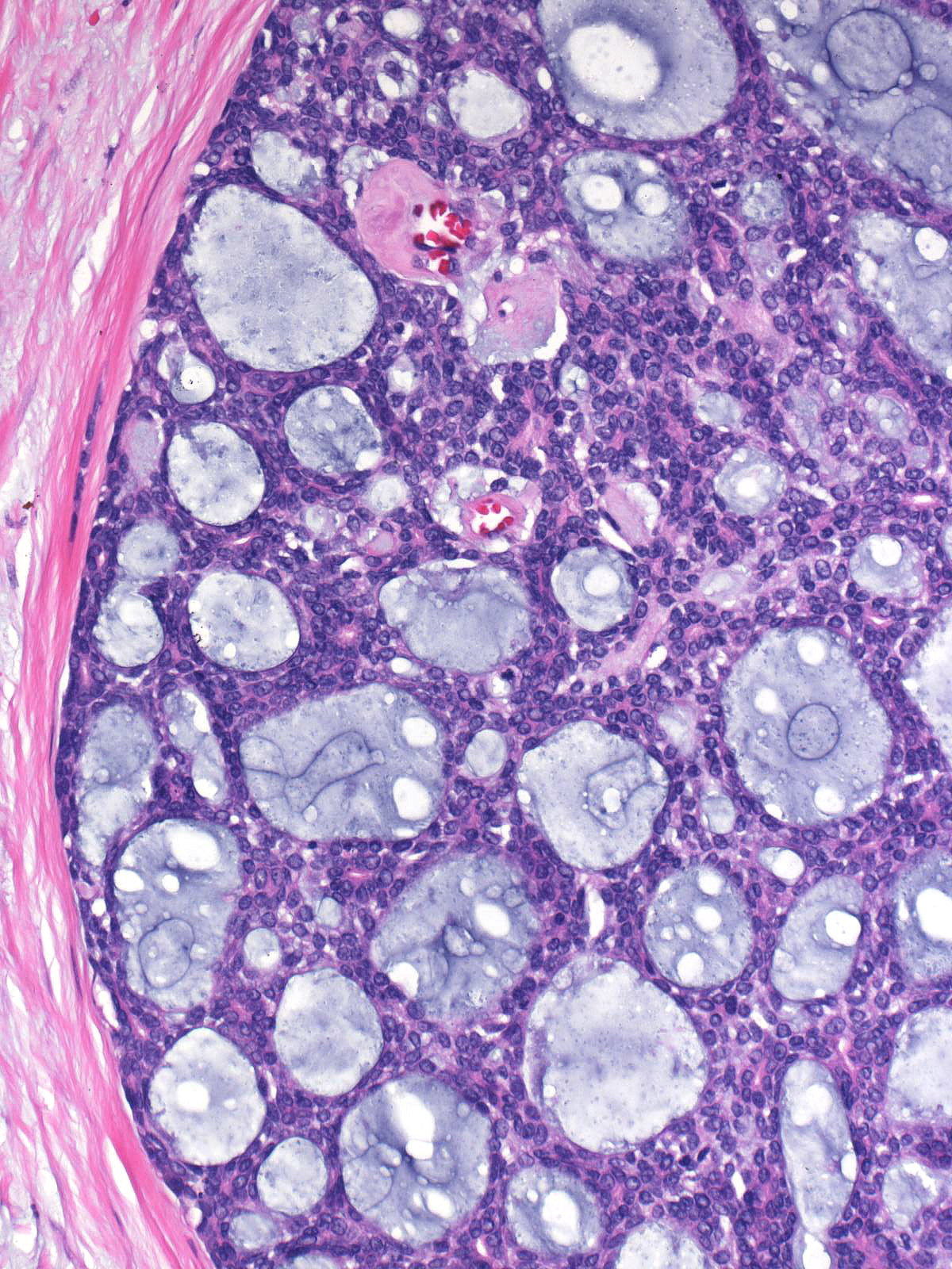
The proportion of luminal and basaloid/myoepithelial cells varies from case to case and from region to region within a single case. Both types of cells create spaces. The luminal cells form small compact tubules, which may appear empty or contain dense, eosinophilic secretion.
 |
 |
| The basaloid/myoepithelial cells produce basement membrane material, which appears pale-blue or pink. |  |
| One can have difficulty distinguishing the two populations of cells in high-grade adenoid cystic carcinomas, but careful histological study and immunohistochemistry will usually allow one to distinguish them. (High-grade adenoid cystic carcinoma) | 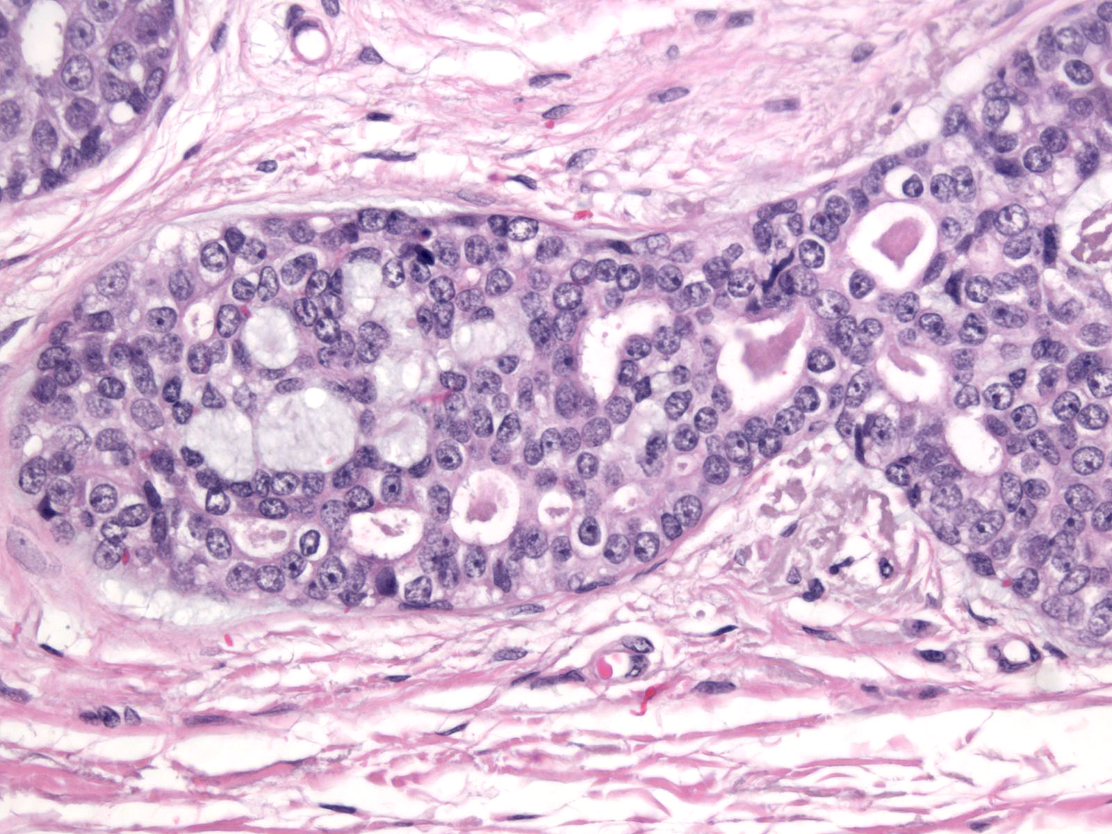 |
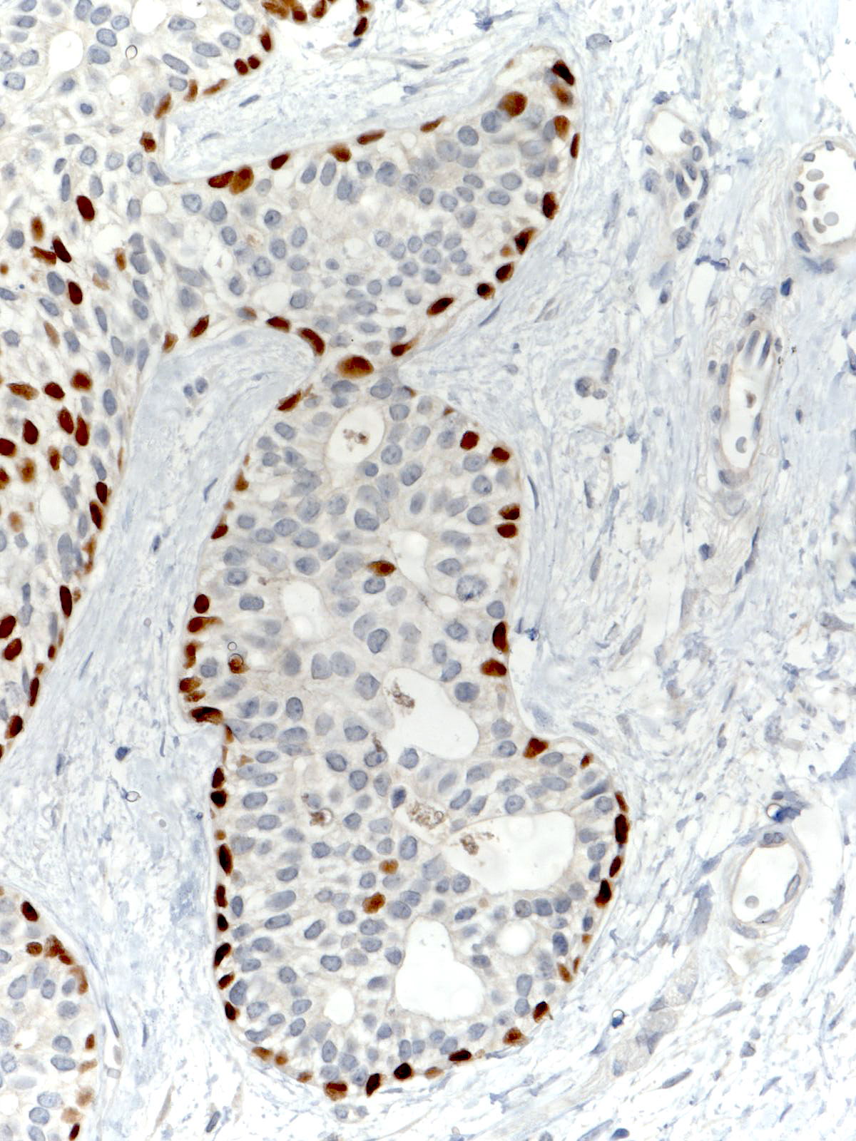
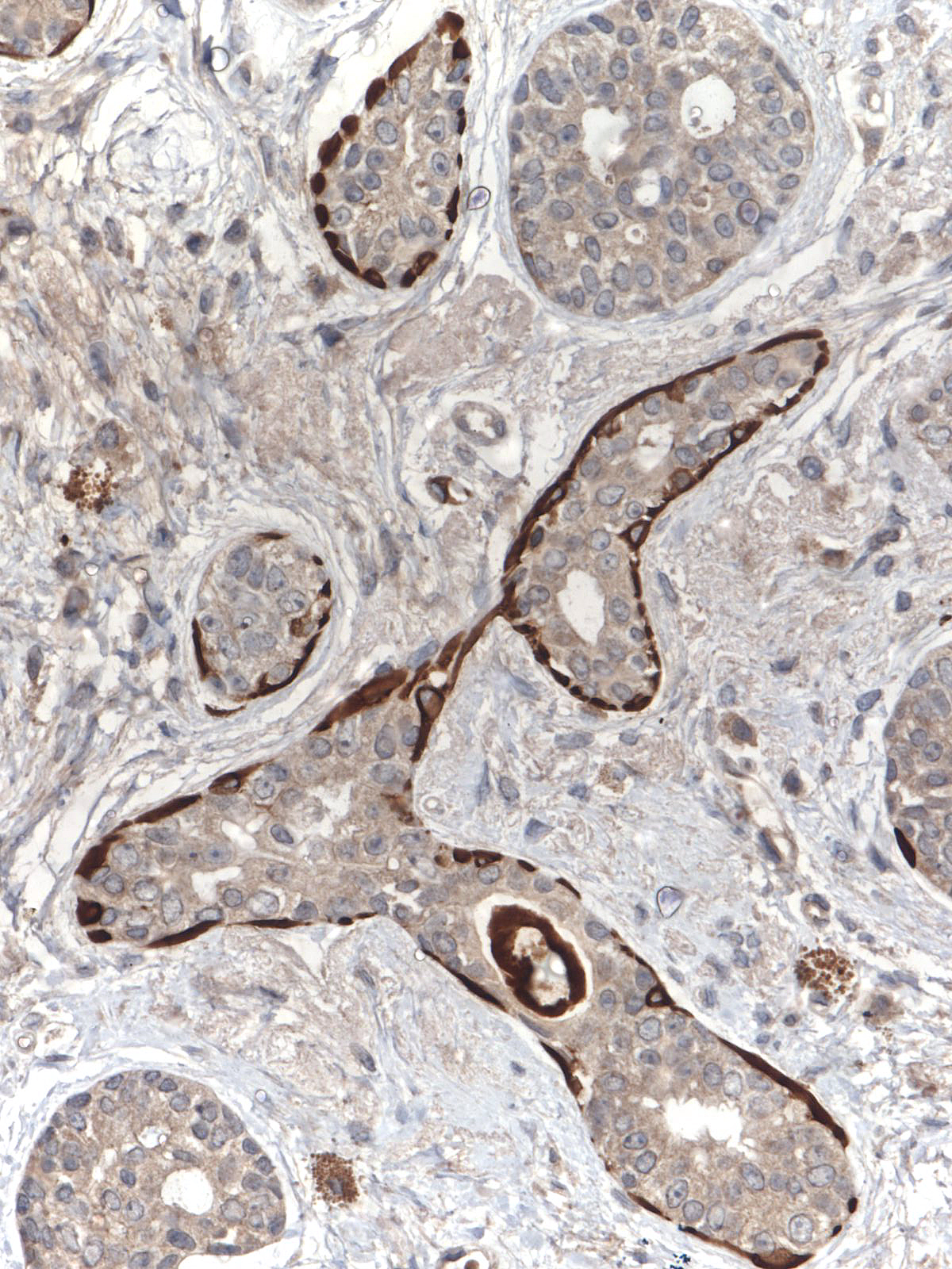
The luminal cells usually demonstrate more intense reactivity for one keratin molecule or another than the basaloid/myoepithelial cells do, and a stain for p63 usually highlights the basaloid/myoepithelial component.
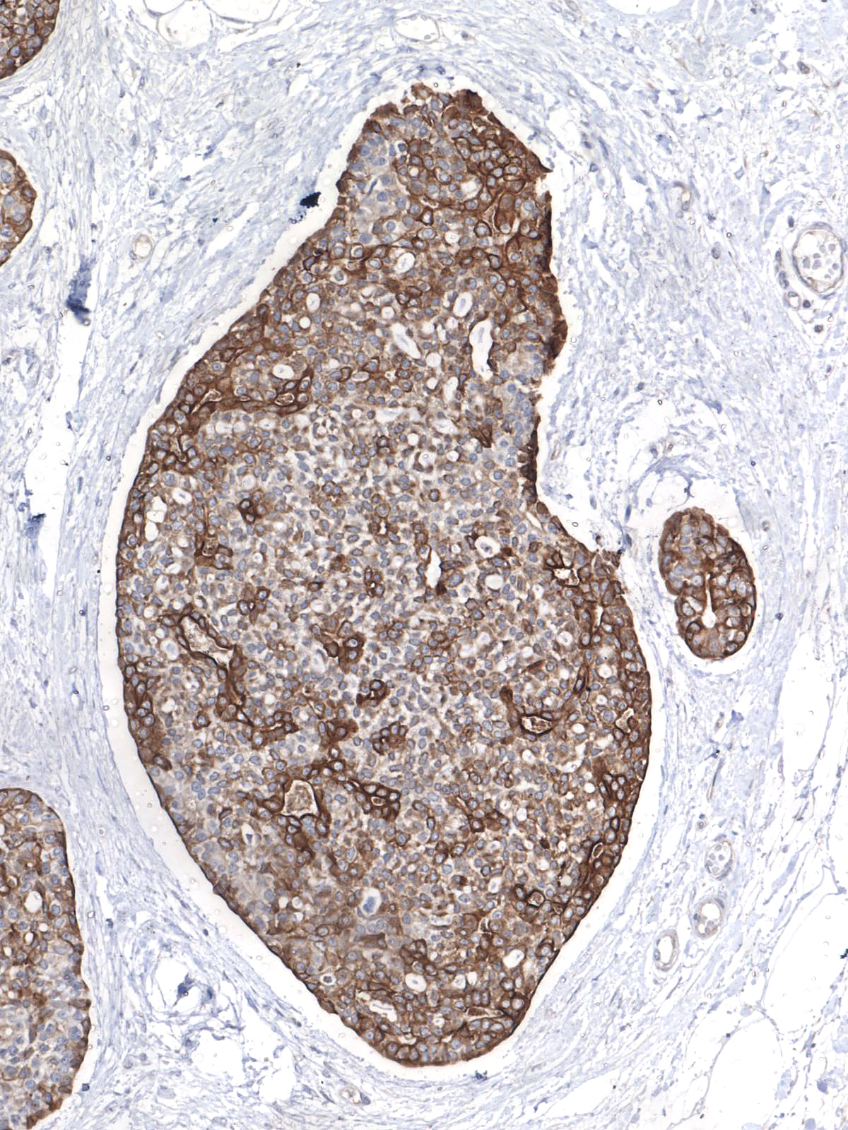 |
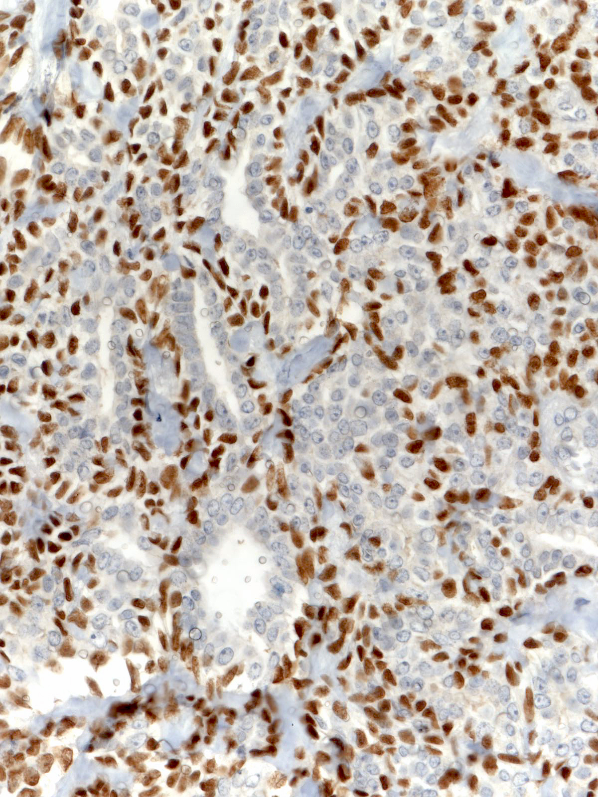 |
Flattening of peripheral basaloid/myoepithelial cells creates an appearance that one could confuse with an in-situ component; however, the infiltrative pattern of growth establishes the invasive nature of the nests.
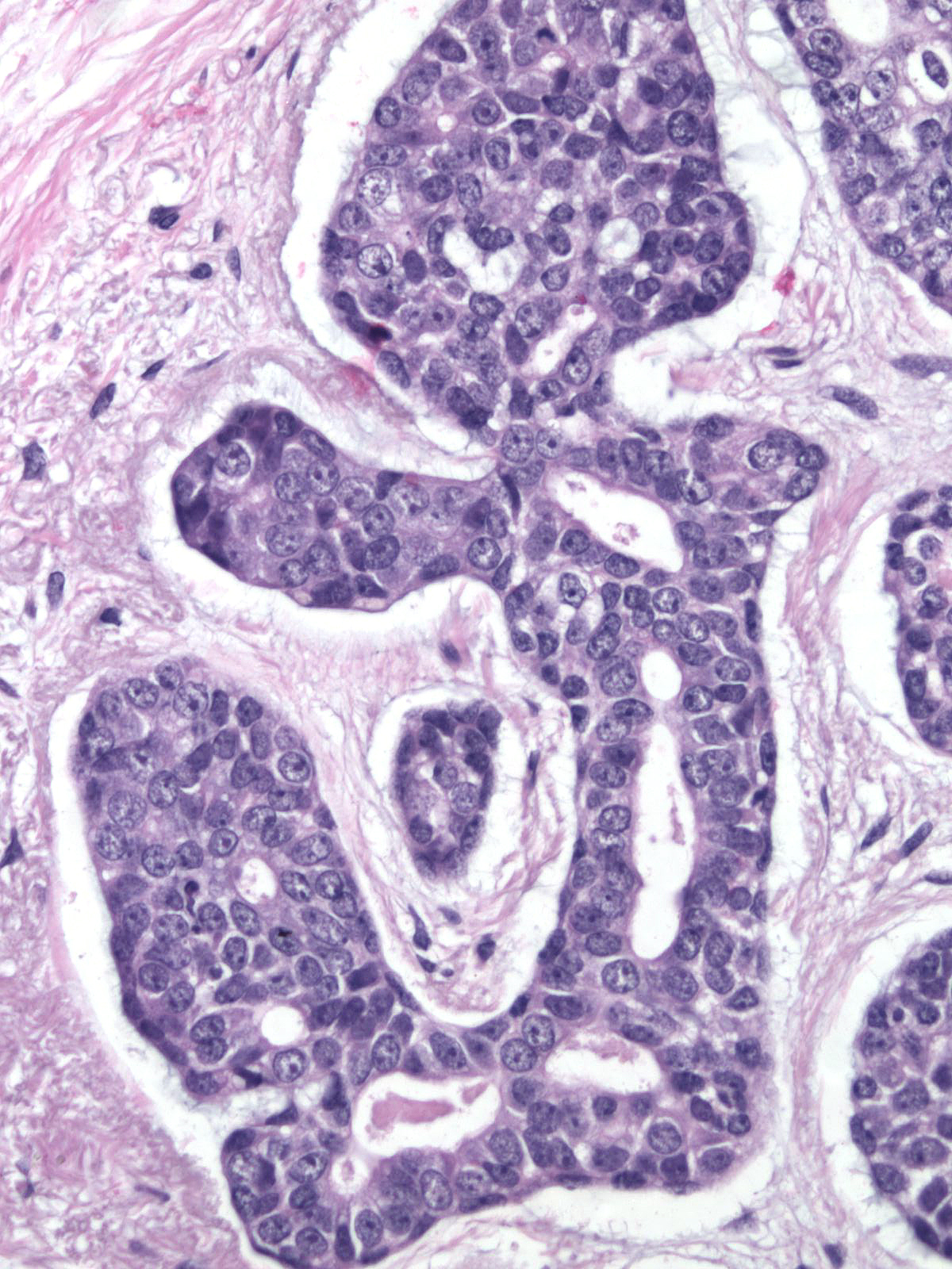 |
 |
 |
| The carcinoma cells in adenoid cystic carcinoma stain for CD117 (c-Kit) and typically do not stain for ER, PR, and HER2. (A stain for CD117 (c-Kit)) | 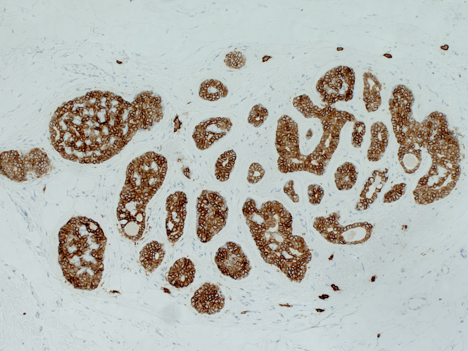 |
tumor) can demonstrate this finding.|24-7 Beta_cat.jpg}}