Difference between revisions of "Breast: DE: Blunt Duct Adenosis"
| Line 15: | Line 15: | ||
{{img1|The stroma of old BDA consists of dense collagen with only a few fibroblasts. Congested capillaries and chronic inflammatory cells no longer stand out prominently.|bda_pic9b.jpg|}} | {{img1|The stroma of old BDA consists of dense collagen with only a few fibroblasts. Congested capillaries and chronic inflammatory cells no longer stand out prominently.|bda_pic9b.jpg|}} | ||
== Quiz == | == Quiz == | ||
| − | <bootstrap_collapse text="Quiz"><p><htmltag tagname="iframe" src="https://hub.partners.org/pathology/assessment/assessment?assessment_id=16564961" height="666" width="100%"></htmltag> | + | <bootstrap_collapse text="Start Quiz"><p><htmltag tagname="iframe" src="https://hub.partners.org/pathology/assessment/assessment?assessment_id=16564961" height="666" width="100%"></htmltag> |
</p></bootstrap_collapse> | </p></bootstrap_collapse> | ||
<hr> | <hr> | ||
<categorytree mode="pages">Breast</categorytree> | <categorytree mode="pages">Breast</categorytree> | ||
[[Category:Breast • Weeks 2 and 3 • Diagnostic Entities]] | [[Category:Breast • Weeks 2 and 3 • Diagnostic Entities]] | ||
Revision as of 05:38, August 28, 2019
Contents
[hide]Introduction
|
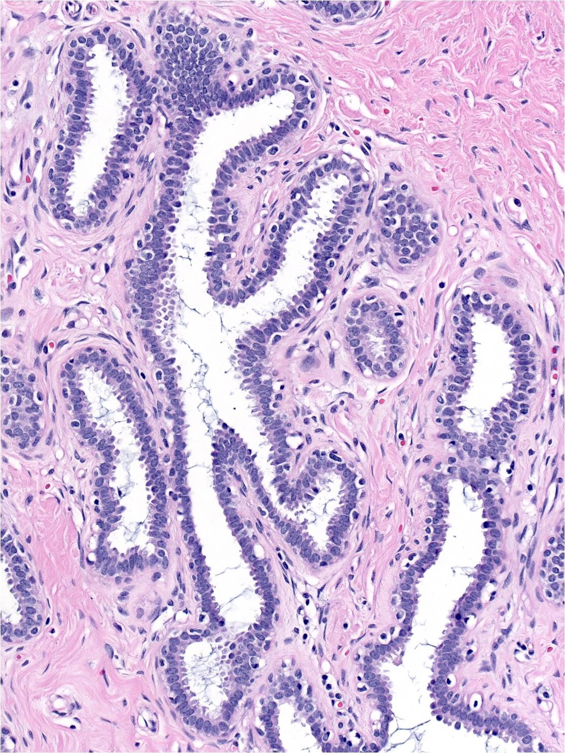
Definition: Blunt duct adenosis (BDA) represents a form of benign lobular hypertrophy in which the proliferative glands look like the ductules of immature lobules. Clinical Significance: BDA has a benign nature and it does not indicate a heightened risk for the development of carcinoma. BDA usually represents an incidental microscopical finding. It does not commonly give rise to clinical signs or symptoms. Calcifications visible on a mammogram can form within the glands, but this happens only rarely. Gross Findings: None Microscopic Findings: The histological features of BDA vary somewhat depending on the age of the focus. Findings common to all examples include: enlargement of the entire lobule; dilatation of the lobular glands, which exhibit a simple branching pattern often referred to as "staghorn-like"; and prominence of the myoepithelial cells. The characteristics of the intralobular (specialized) stroma varies with the age of the focus. The specialized stroma often appears cellular, and the extracellular matrix contains collagen or myxoid ground substances. Numerous lymphocytes and plasma cells may be present. Differential Diagnosis: Early usual ductal hyperplasia (glands usually lack the simple branching pattern and the myoepithelial cell layer is less prominent); flat epithelial atypia (low-grade cytologic atypia, orderly arrangement, non-prominent myoepithelial cells). Discussion: |
 |
Early Phase BDA
| Early foci of blunt duct adenosis stand out clearly from the surrounding breast parenchyma. The glands look like blunt tubules with a simple branching pattern. Early pathologists were reminded of the the simple branching pattern of the antlers of male deer (stags) when observing the glands of BDA. The myoepithelial cell layer and specialized stroma are also especially prominent in early BDA. | 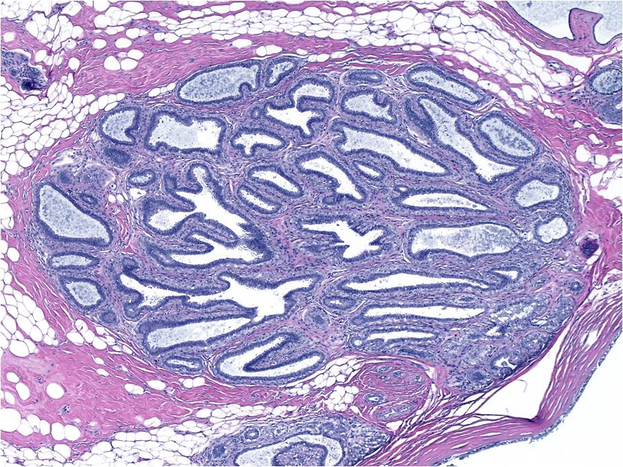 |
| The epithelium lining the glands consists of a layer of closely packed columnar cells overlying a layer of prominent myoepithelial cells. The intralobular (specialized) stroma consists of many fibroblasts embedded in an extracellular matrix. The matrix usually consists of collagen but may contain myxoid ground substances. | 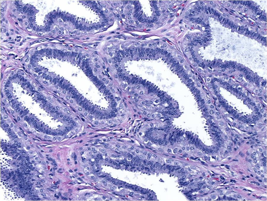 |
| The luminal cells possess oval nuclei, granular chromatin, easily seen nucleoli, and dense eosinophilic apical cytoplasm. The luminal cell nuclei tend to sit at the bases of the cells; however, when the cells become especially crowded, stratification and overlapping of nuclei can occur. The myoepithelial cells form a continuous layer beneath the luminal cells. The myoepithelial cells have oval or round nuclei, pale chromatin, small nucleoli, and abundant pale or clear cytoplasm. The cytoplasm of each myoepithelial cell seems to touch the cytoplasm of its neighbor. | 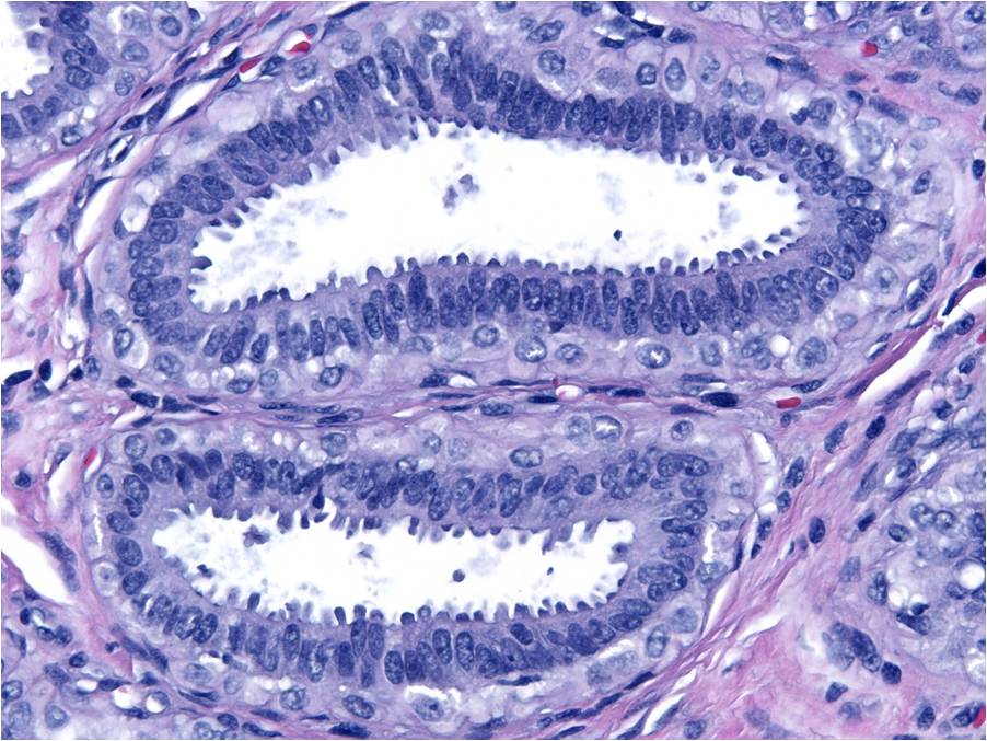 |
| A stain for p63 will highlight the prominent myoepithelial cell population. | 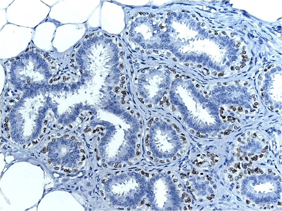 |
| Small amounts of pale blue, myxoid material may be seen in addition to collagen in the stroma. The presence of congested capillaries, lymphocytes, and plasma cells characterize certain cases. | 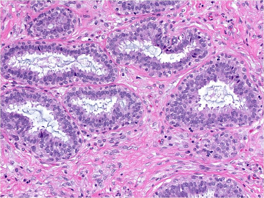 |
| Proliferation of the epithelial cells within blunt duct adenosis may create small foci of usual ductal hyperplasia. | 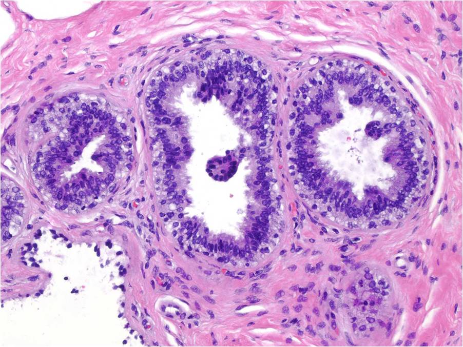 |
Late Phase BDA
| Abatement of proliferative features characterizes late phase BDA. Here we see BDA intermediate between early and late phases. The large size of the lobules persists; however, the glands appear more widely spaced as the stroma loses some of its cellularity, and the epithelium less lush. | 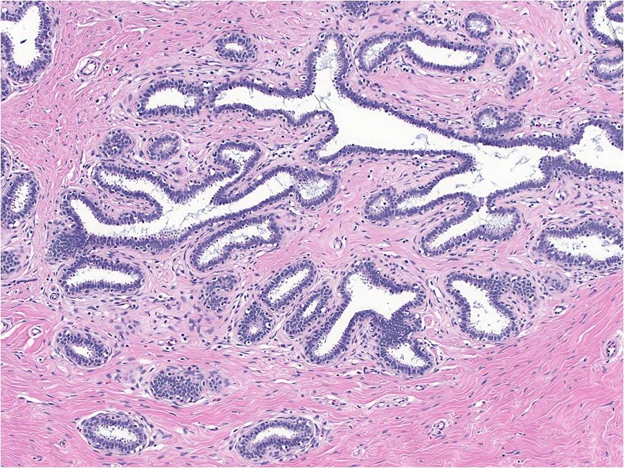 |
| Although the glands in older BDA still demonstrate simple branching, they take on rounder contours. In this picture we see true late phase BDA. The glands have rounder contours and are widely spaced, and the stroma has lost most of its cellularity. | 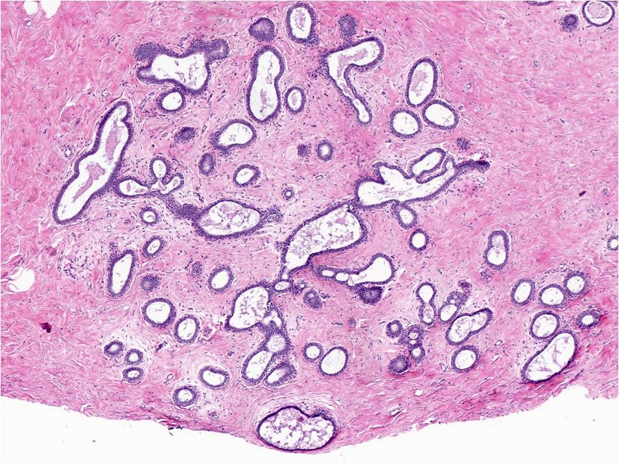 |
| Close examination reveals that the epithelium lining the glands appears less lush and that certain of the once columnar luminal cells have become cuboidal. The nuclei still possess oval to round nuclei, granular chromatin, small nucleoli, and modest amounts of apical cytoplasm. Although the myoepithelial cells still form a noticeable layer, they appear smaller and they contain small, dark nuclei and scant cytoplasm. | 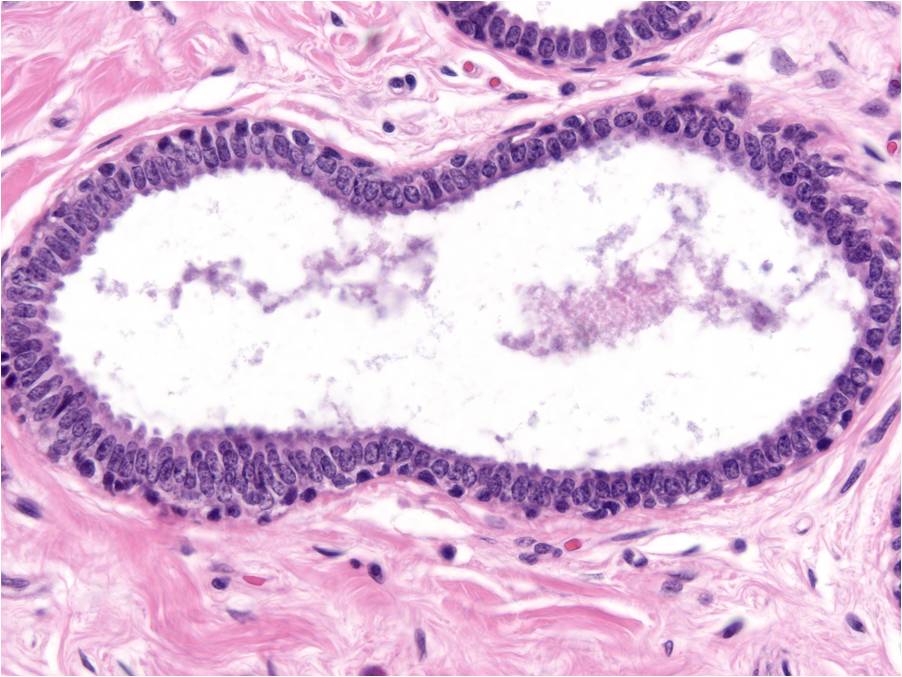 |
| The stroma of old BDA consists of dense collagen with only a few fibroblasts. Congested capillaries and chronic inflammatory cells no longer stand out prominently. | 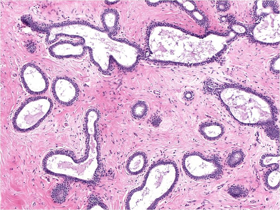 |
Quiz