Difference between revisions of "Pseudoangiomatous Stromal Hyperplasia (PASH)"
| (2 intermediate revisions by the same user not shown) | |||
| Line 3: | Line 3: | ||
<br> | <br> | ||
== Introduction == | == Introduction == | ||
| − | {{dxintro|Pseudoangiomatous Stromal Hyperplasia (PASH)|Pseudoangiomatous stromal hyperplasia (PASH) represents a proliferation of myofibroblasts of the nonspecialized stroma.|Most examples represent incidental findings detected during microscopic examination. Rarely, the lesion can give rise to palpable nodules, which demonstrate clinical features usually suggestive of a benign mass. Regions of pseudoangiomatous stromal hyperplasia can also create densities on mammograms and enhancing areas on MRI. All forms of pseudoangiomatous stromal hyperplasia have an entirely benign nature.|Macroscopic examination will not reveal small foci of PASH. Larger, clinically evident foci appear as well defined, homogenous, white, rubbery nodules.|The proliferative myofibroblasts usually form an interlacing network of channels that resemble blood vessels. In a form known as fascicular pseudoangiomatous stromal hyperplasia, the myofibroblasts grow in compact bundles rather than channels. The edges of most foci of PASH blend with the surrounding parenchyma; however, occasional examples form well defined masses, which pathologists refer to as nodular pseudoangiomatous stromal hyperplasia. The proliferation of myofibroblasts is usually accompanied by expansion of the specialized stroma, simplification of the glandular branching pattern, and proliferaton of the glandular epithelium. | + | {{dxintro|Pseudoangiomatous Stromal Hyperplasia (PASH)|Pseudoangiomatous stromal hyperplasia (PASH) represents a proliferation of myofibroblasts of the nonspecialized stroma.|Most examples represent incidental findings detected during microscopic examination. Rarely, the lesion can give rise to palpable nodules, which demonstrate clinical features usually suggestive of a benign mass. Regions of pseudoangiomatous stromal hyperplasia can also create densities on mammograms and enhancing areas on MRI. All forms of pseudoangiomatous stromal hyperplasia have an entirely benign nature.|Macroscopic examination will not reveal small foci of PASH. Larger, clinically evident foci appear as well defined, homogenous, white, rubbery nodules.|The proliferative myofibroblasts usually form an interlacing network of channels that resemble blood vessels. In a form known as fascicular pseudoangiomatous stromal hyperplasia, the myofibroblasts grow in compact bundles rather than channels. The edges of most foci of PASH blend with the surrounding parenchyma; however, occasional examples form well defined masses, which pathologists refer to as nodular pseudoangiomatous stromal hyperplasia. The proliferation of myofibroblasts is usually accompanied by expansion of the specialized stroma, simplification of the glandular branching pattern, and proliferaton of the glandular epithelium.|Low-grade angiosarcoma (true vascular spaces that infiltrate specialized stroma and lobules), fibrocystic changes (lacks pseudoangiomatous change), myofibroblastoma (lacks glands within the mass)|An orderly proliferation of myofibroblasts of the nonspecialized stroma constitutes the defining microscopical feature of pseudoangiomatous stromal hyperplasia.|35-1 PASH_-_intro.jpg|PASH consists of a proliferation of myofibroblasts in the nonspecialized stroma. Entrapped glands and specialized stroma usually appear hyperplastic.}} |
A typical example of PASH demonstrates a network of myofibroblasts occupying the nonspecialized stroma. | A typical example of PASH demonstrates a network of myofibroblasts occupying the nonspecialized stroma. | ||
Latest revision as of 08:52, July 17, 2020
Contents
Introduction
|
Definition: Pseudoangiomatous stromal hyperplasia (PASH) represents a proliferation of myofibroblasts of the nonspecialized stroma. Clinical Significance: Most examples represent incidental findings detected during microscopic examination. Rarely, the lesion can give rise to palpable nodules, which demonstrate clinical features usually suggestive of a benign mass. Regions of pseudoangiomatous stromal hyperplasia can also create densities on mammograms and enhancing areas on MRI. All forms of pseudoangiomatous stromal hyperplasia have an entirely benign nature. Gross Findings: Macroscopic examination will not reveal small foci of PASH. Larger, clinically evident foci appear as well defined, homogenous, white, rubbery nodules. Microscopic Findings: The proliferative myofibroblasts usually form an interlacing network of channels that resemble blood vessels. In a form known as fascicular pseudoangiomatous stromal hyperplasia, the myofibroblasts grow in compact bundles rather than channels. The edges of most foci of PASH blend with the surrounding parenchyma; however, occasional examples form well defined masses, which pathologists refer to as nodular pseudoangiomatous stromal hyperplasia. The proliferation of myofibroblasts is usually accompanied by expansion of the specialized stroma, simplification of the glandular branching pattern, and proliferaton of the glandular epithelium. Differential Diagnosis: Low-grade angiosarcoma (true vascular spaces that infiltrate specialized stroma and lobules), fibrocystic changes (lacks pseudoangiomatous change), myofibroblastoma (lacks glands within the mass) Discussion: An orderly proliferation of myofibroblasts of the nonspecialized stroma constitutes the defining microscopical feature of pseudoangiomatous stromal hyperplasia. |
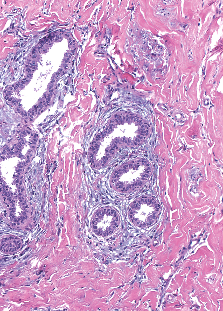 |
A typical example of PASH demonstrates a network of myofibroblasts occupying the nonspecialized stroma.
 |
 |
| In this example of fascicular PASH, the myofibroblasts form bundles. |  |
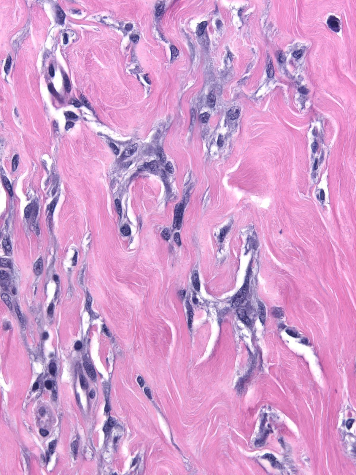
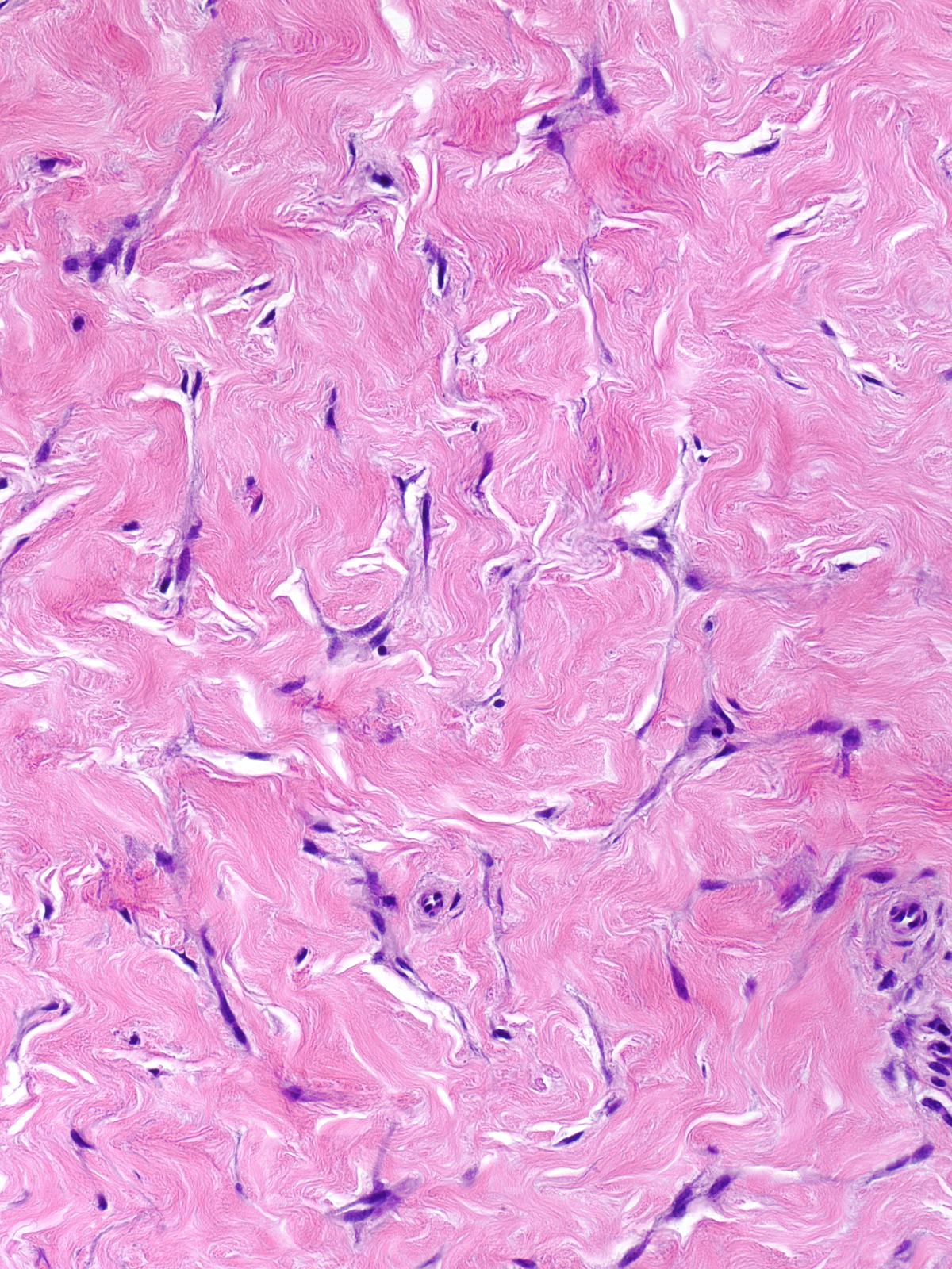
In classic, well-established cases, the spindle shaped cells possess small amounts of cytoplasm, are spindled in shape, have ill-defined cell boarders, and contain small, oval to spindly, hyperchromatic nuclei and inconspicuous nucleoli. The cytoplasm stains pale blue to grey. The cells do not display mitotic figures. They stain for CD34, but they do not stain for CD31 or other proteins found in endothelial cells. Other cytological features vary according to the age of the focus.
 |
 |
| In cellular PASH, the myofibroblasts sometimes contain modest amounts of cytoplasm and oval nuclei containing granular chromatin and a single small nucleolus. Such cellular lesions may lack slit-like spaces. | 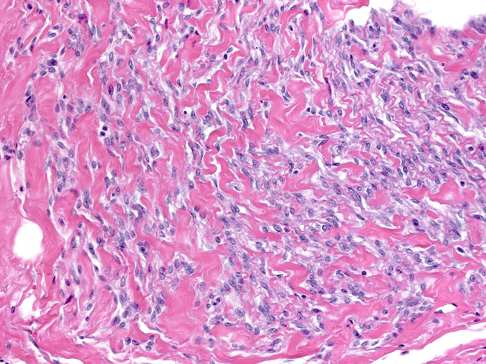 |
| Foci of PASH usually have an indistinct border. The myofibroblasts typically trail off into the surrounding parenchyma and sometimes into the fat. | 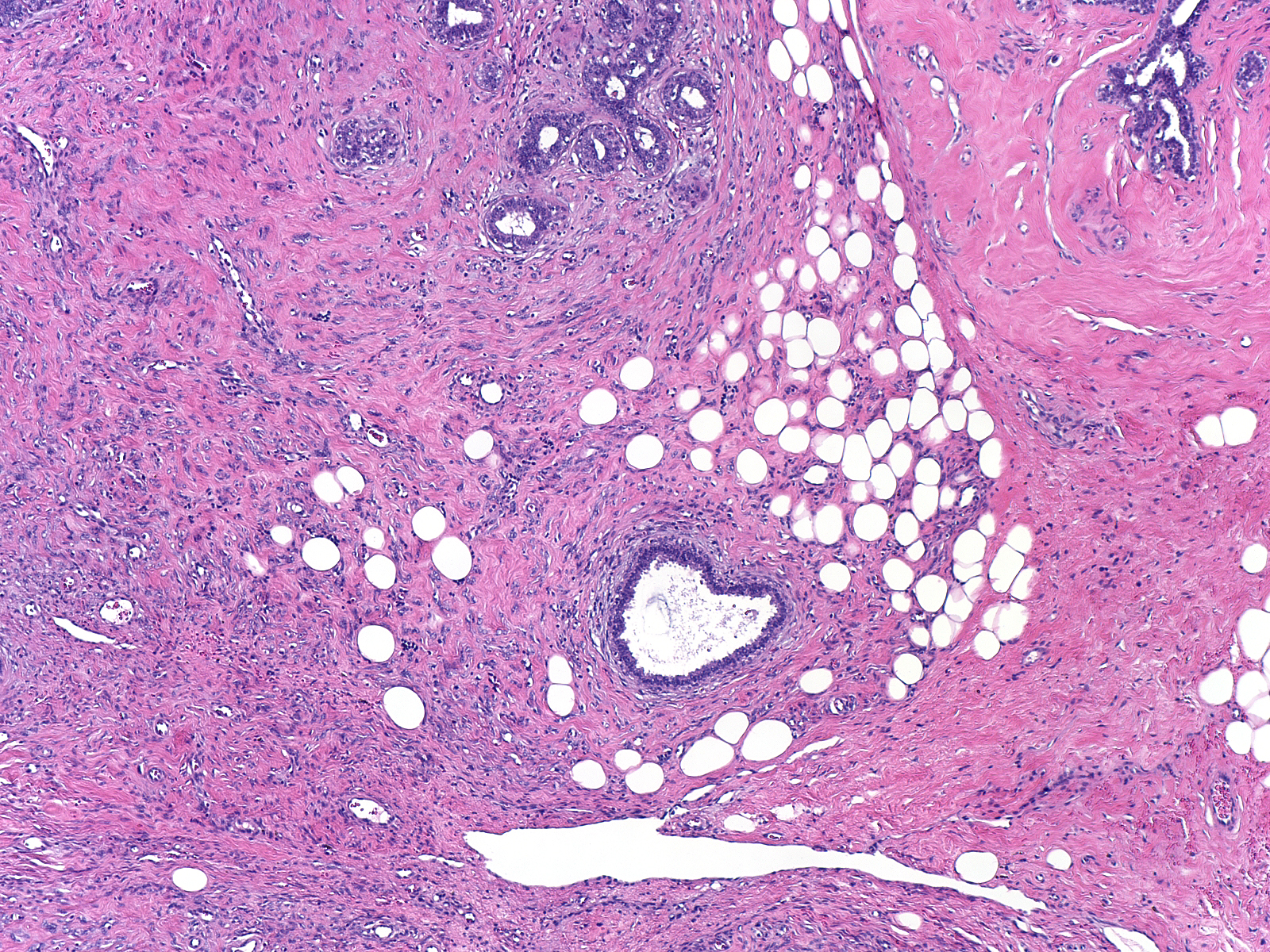 |
| Masses classified as nodular pseudoangiomatous stromal hyperplasia, have well-defined borders. These lesions may represent hamartomas with superimposed PASH. |  |
Glandular tissue surrounded by pseudoangiomatous stromal hyperplasia demonstrates proliferative changes. Entrapped lobules appear larger than uninvolved ones and their acini less closely packed.
 |
 |
The glands exhibit a tubular configuration and a simple branching pattern. Expanded zones of specialized stroma encase the acini and ductules in uniform cuffs consisting of abundant fibroblasts and myxoid ground substances. Proliferative epithelium lines the glands. The epithelial cells usually grow in a single layer, but they can show marked stratification.
 |
 |
| A subtle proliferation of true small blood vessels usually accompanies the proliferation of myofibroblasts. | 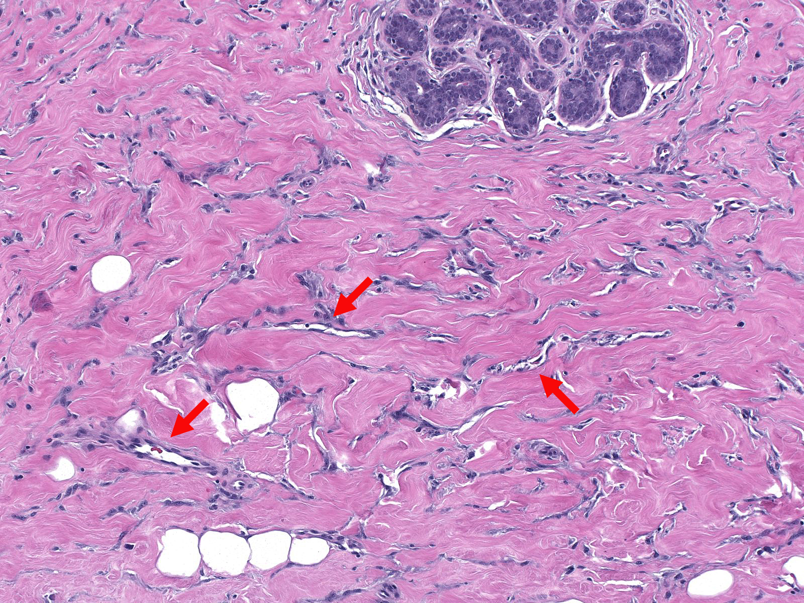 |