Difference between revisions of "Phyllodes Tumor"
(Created page with "__NOCACHE__{{DISPLAYTITLE:Phyllodes Tumor}} {{:TOC}} == General == Image:33-1 Gross_1_-_CROP.jpg|thumb|The cut surface of a phyllodes tumor can show hemorrhage, fronds of st...") |
|||
| (2 intermediate revisions by 2 users not shown) | |||
| Line 1: | Line 1: | ||
| − | __NOCACHE__ | + | __NOCACHE__ |
{{:TOC}} | {{:TOC}} | ||
| − | == | + | <br> |
| − | + | == Introduction == | |
| − | + | {{dxintro|Phyllodes Tumor|Phyllodes tumors are sarcomas derived from cells of the specialized stroma.|Phyllodes tumors present as masses, which often have cystic regions. If shelled out, these tumors can recur, although they do so in only a minority of cases. With the development of several recurrences, the grade of the tumor usually increases. For this reason, surgeons usually strive to excise a modest rim of uninvolved tissue around a phyllodes tumor. This approach may require additional surgery after the diagnostic excision. Phyllodes tumors are capable of metastasizing. When they do so, phyllodes tumors spread in the distribution characteristic of a sarcoma, and the metastatic nodules consist only of stromal cells.|Phyllodes tumors typically form a well defined, round, fleshy, tan mass, which typically contains spaces into which fronds of neoplastic tissue project.|Neoplastic stromal cells and entrapped, distorted, pre-existing ducts and lobules make up most phyllodes tumors. The two components usually mingle throughout the mass; however, in rare tumors, the stromal cells overgrow the glandular elements and make it difficult to detect them.|Conventional sarcoma (no glandular component), spindle cell carcinoma (malignant glandular element, keratin positivity)|Like fibroadenomas, phyllodes tumors originate from specialized stromal cells; however, the properties of the neoplastic cells in the two lesions differ. Those in phyllodes tumors do not retain the orderly relationship and organization with respect to the glandular elements that characterizes the stromal cells of fibroadenomas.|33-1 Gross_1_-_CROP.jpg|The cut surface of a phyllodes tumor can show hemorrhage, fronds of stroma, and cyst formation. Originally called cystosarcoma phyllodes, its name comes from the Greek words sarcoma (fleshy tumor) and phyllon (leaf).}} | |
| − | |||
| − | |||
| − | |||
| − | |||
| − | |||
| − | |||
| − | |||
| − | |||
| − | |||
| − | |||
Histological findings that reflect this lack of organization include: regional variation in the structure of the tumor, variation in the density of the stromal cells, large collections of stromal cells lacking glands, and fascicles of stromal cells growing without orientation to the glands. The stromal cells often display other commonly cited features such as dense cellularity, atypia, and mitotic activity, but one need not observe these findings to make the diagnosis of phyllodes tumor. | Histological findings that reflect this lack of organization include: regional variation in the structure of the tumor, variation in the density of the stromal cells, large collections of stromal cells lacking glands, and fascicles of stromal cells growing without orientation to the glands. The stromal cells often display other commonly cited features such as dense cellularity, atypia, and mitotic activity, but one need not observe these findings to make the diagnosis of phyllodes tumor. | ||
{{img4|33-2 Variation_1.jpg|33-3 Variation_2.jpg|The neoplastic cells of a phyllodes tumors disrupt the orderly arrangement of normal breast tissue and display regional variations in structure and cellularity.|}} | {{img4|33-2 Variation_1.jpg|33-3 Variation_2.jpg|The neoplastic cells of a phyllodes tumors disrupt the orderly arrangement of normal breast tissue and display regional variations in structure and cellularity.|}} | ||
| Line 34: | Line 24: | ||
{{img4|33-19 Invasion_1.jpg|33-20 Invasion_2.jpg||}} | {{img4|33-19 Invasion_1.jpg|33-20 Invasion_2.jpg||}} | ||
{{img1|The epithelium of the entrapped glands usually demonstrates evidence of proliferation such as cellular and nuclear enlargement, mitotic activity of the epithelial cells, and prominence of the myoepithelial cells.|33-21 Epithelium-3.jpg}} | {{img1|The epithelium of the entrapped glands usually demonstrates evidence of proliferation such as cellular and nuclear enlargement, mitotic activity of the epithelial cells, and prominence of the myoepithelial cells.|33-21 Epithelium-3.jpg}} | ||
| − | Compare the attenuated epithelium of the fibroadenoma on the left with the proliferative epithelium of the phyllodes tumor on the right. | + | {{img1|Compare the attenuated epithelium of the fibroadenoma on the left with the proliferative epithelium of the phyllodes tumor on the right.|33-22 Composite_2.jpg}} |
| − | |||
{{img1|Growth of the epithelium in a flat pattern causes the glands to dilate and to assume unusual shapes likened to those of animals.|33-24 Dilated.jpg}} | {{img1|Growth of the epithelium in a flat pattern causes the glands to dilate and to assume unusual shapes likened to those of animals.|33-24 Dilated.jpg}} | ||
{{img1|The epithelium can give rise to ductal hyperplasia, which can appear especially florid.|33-25 UDH-4.jpg}} | {{img1|The epithelium can give rise to ductal hyperplasia, which can appear especially florid.|33-25 UDH-4.jpg}} | ||
| Line 48: | Line 37: | ||
Phyllodes tumors usually arise from pre-existing fibroepithelial tumors. Fibroadenomas probably represent the most common lesion of origin, but phyllodes tumors can also arise from hamartomas. The presence of such an underlying fibroepithelial tumor contributes to the regional variability in glandular architecture, stromal cellularity, and composition of the extracellular matrix that characterize phyllodes tumors. | Phyllodes tumors usually arise from pre-existing fibroepithelial tumors. Fibroadenomas probably represent the most common lesion of origin, but phyllodes tumors can also arise from hamartomas. The presence of such an underlying fibroepithelial tumor contributes to the regional variability in glandular architecture, stromal cellularity, and composition of the extracellular matrix that characterize phyllodes tumors. | ||
{{img4|33-31 PT_FBA.jpg|33-32 PT_ham.jpg|Phyllodes tumor arising in a fibroadenoma|Phyllodes tumor arising in a hamartoma}} | {{img4|33-31 PT_FBA.jpg|33-32 PT_ham.jpg|Phyllodes tumor arising in a fibroadenoma|Phyllodes tumor arising in a hamartoma}} | ||
| − | + | {{:mgh:breast-footer}} | |
| − | |||
| − | |||
Latest revision as of 08:48, July 17, 2020
Contents
Introduction
|
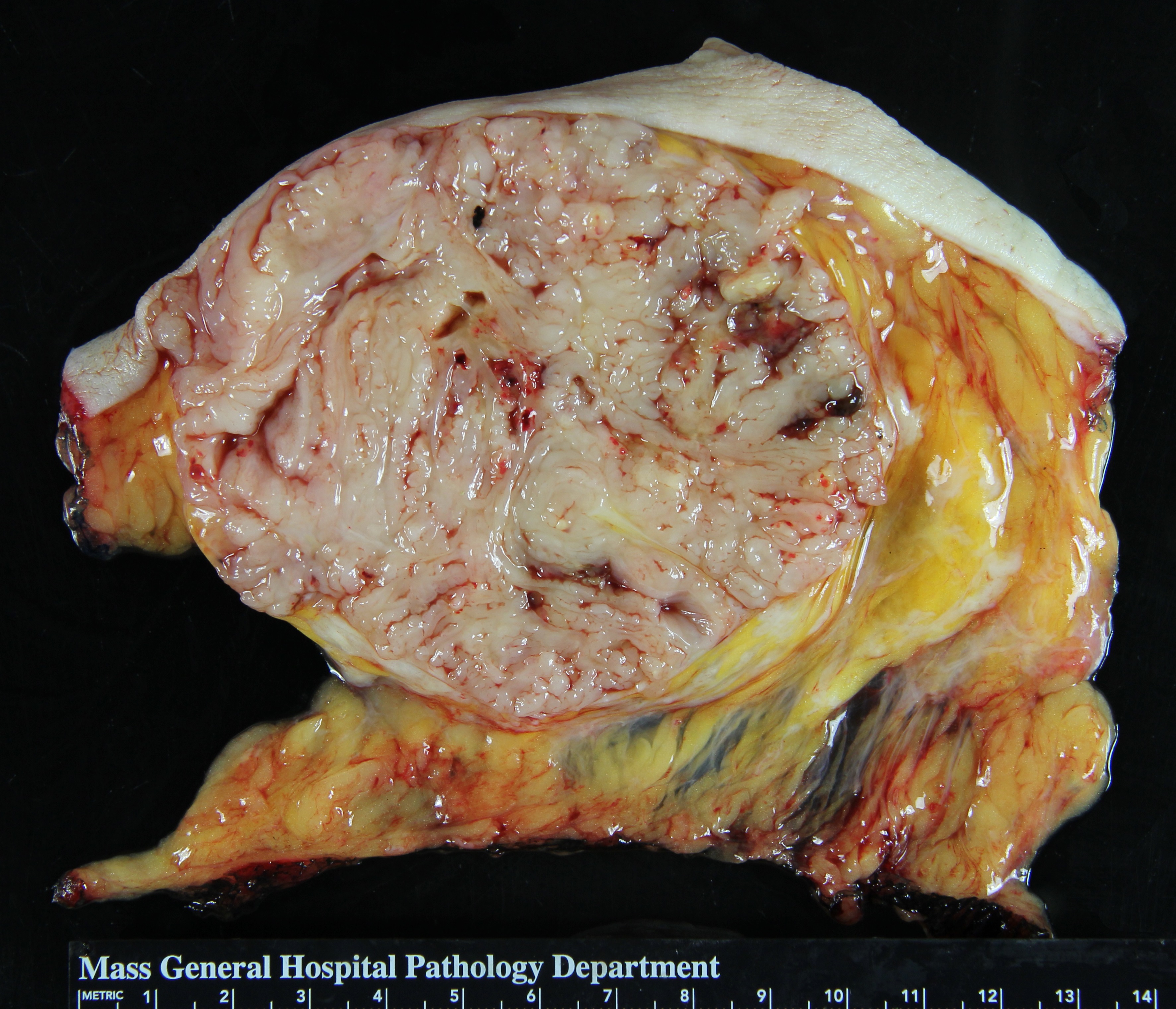
Definition: Phyllodes tumors are sarcomas derived from cells of the specialized stroma. Clinical Significance: Phyllodes tumors present as masses, which often have cystic regions. If shelled out, these tumors can recur, although they do so in only a minority of cases. With the development of several recurrences, the grade of the tumor usually increases. For this reason, surgeons usually strive to excise a modest rim of uninvolved tissue around a phyllodes tumor. This approach may require additional surgery after the diagnostic excision. Phyllodes tumors are capable of metastasizing. When they do so, phyllodes tumors spread in the distribution characteristic of a sarcoma, and the metastatic nodules consist only of stromal cells. Gross Findings: Phyllodes tumors typically form a well defined, round, fleshy, tan mass, which typically contains spaces into which fronds of neoplastic tissue project. Microscopic Findings: Neoplastic stromal cells and entrapped, distorted, pre-existing ducts and lobules make up most phyllodes tumors. The two components usually mingle throughout the mass; however, in rare tumors, the stromal cells overgrow the glandular elements and make it difficult to detect them. Differential Diagnosis: Conventional sarcoma (no glandular component), spindle cell carcinoma (malignant glandular element, keratin positivity) Discussion: Like fibroadenomas, phyllodes tumors originate from specialized stromal cells; however, the properties of the neoplastic cells in the two lesions differ. Those in phyllodes tumors do not retain the orderly relationship and organization with respect to the glandular elements that characterizes the stromal cells of fibroadenomas. |
 |
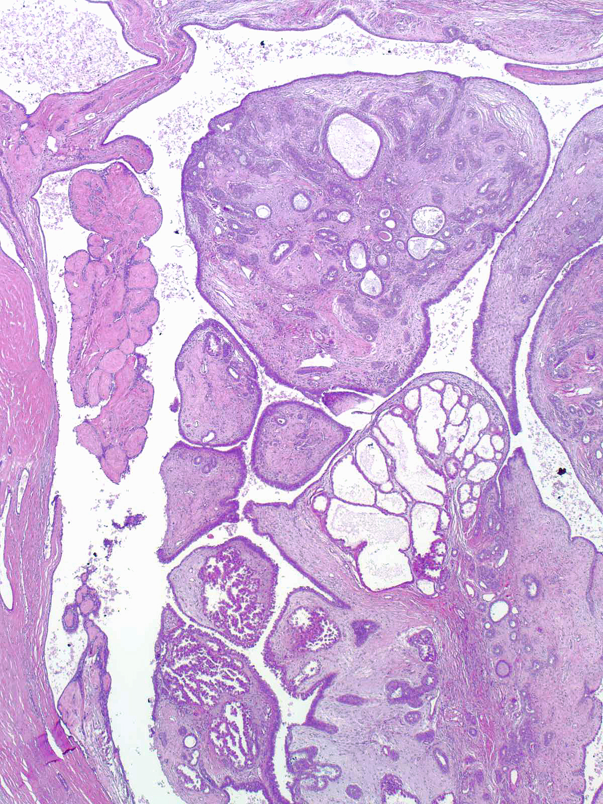
Histological findings that reflect this lack of organization include: regional variation in the structure of the tumor, variation in the density of the stromal cells, large collections of stromal cells lacking glands, and fascicles of stromal cells growing without orientation to the glands. The stromal cells often display other commonly cited features such as dense cellularity, atypia, and mitotic activity, but one need not observe these findings to make the diagnosis of phyllodes tumor.
 |
 |
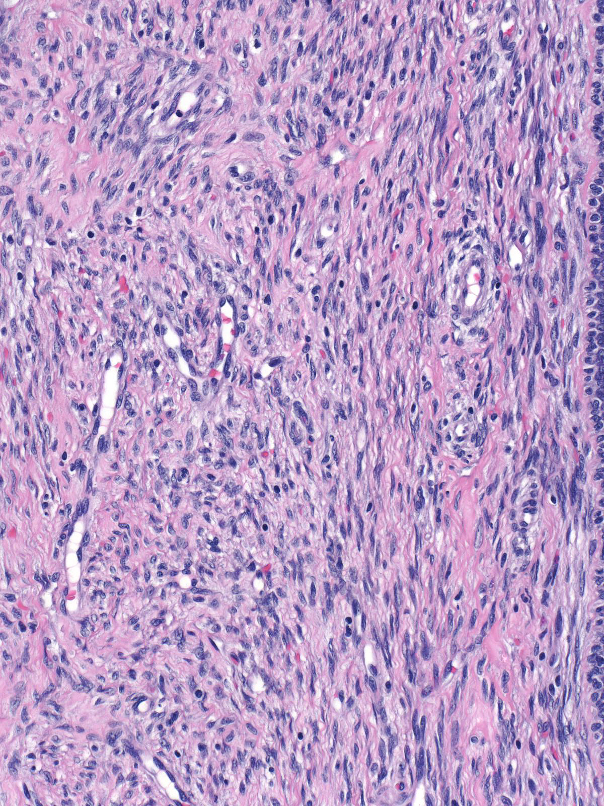
The neoplastic cells of a phyllodes tumors disrupt the orderly arrangement of normal breast tissue and display regional variations in structure and cellularity.
 |
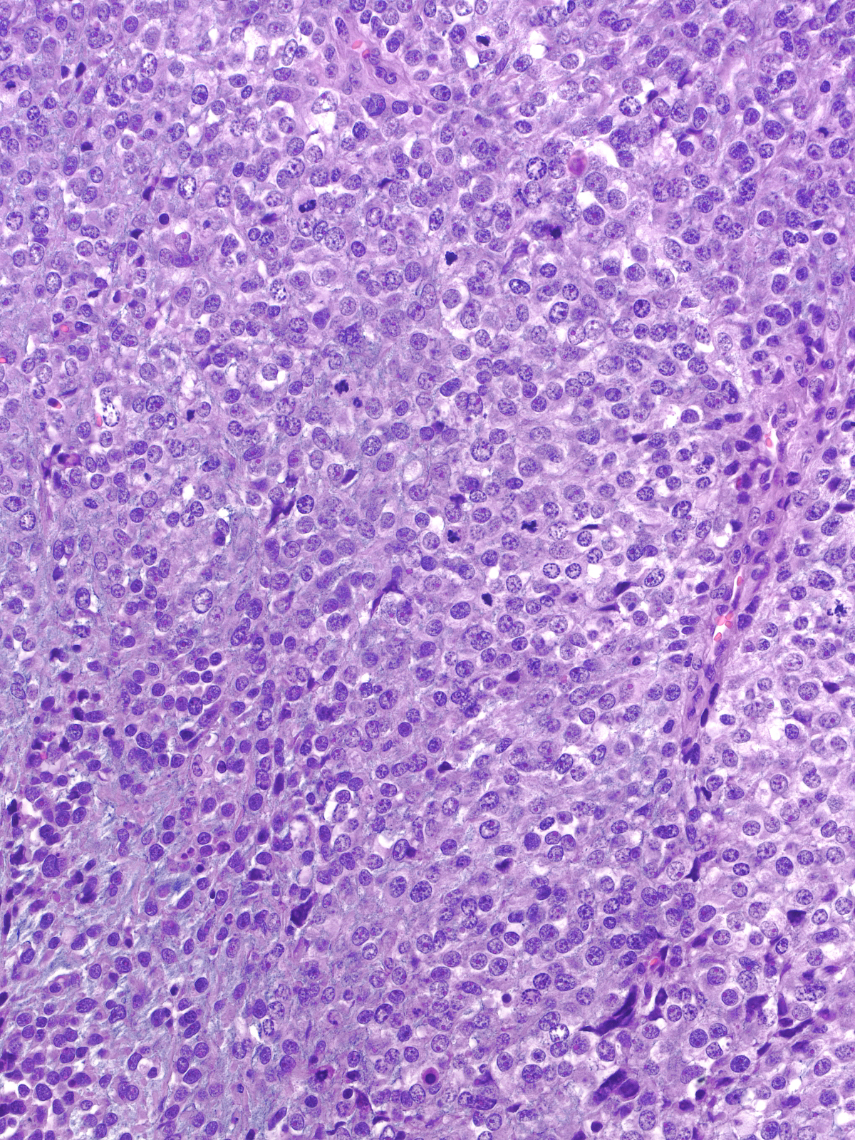 |
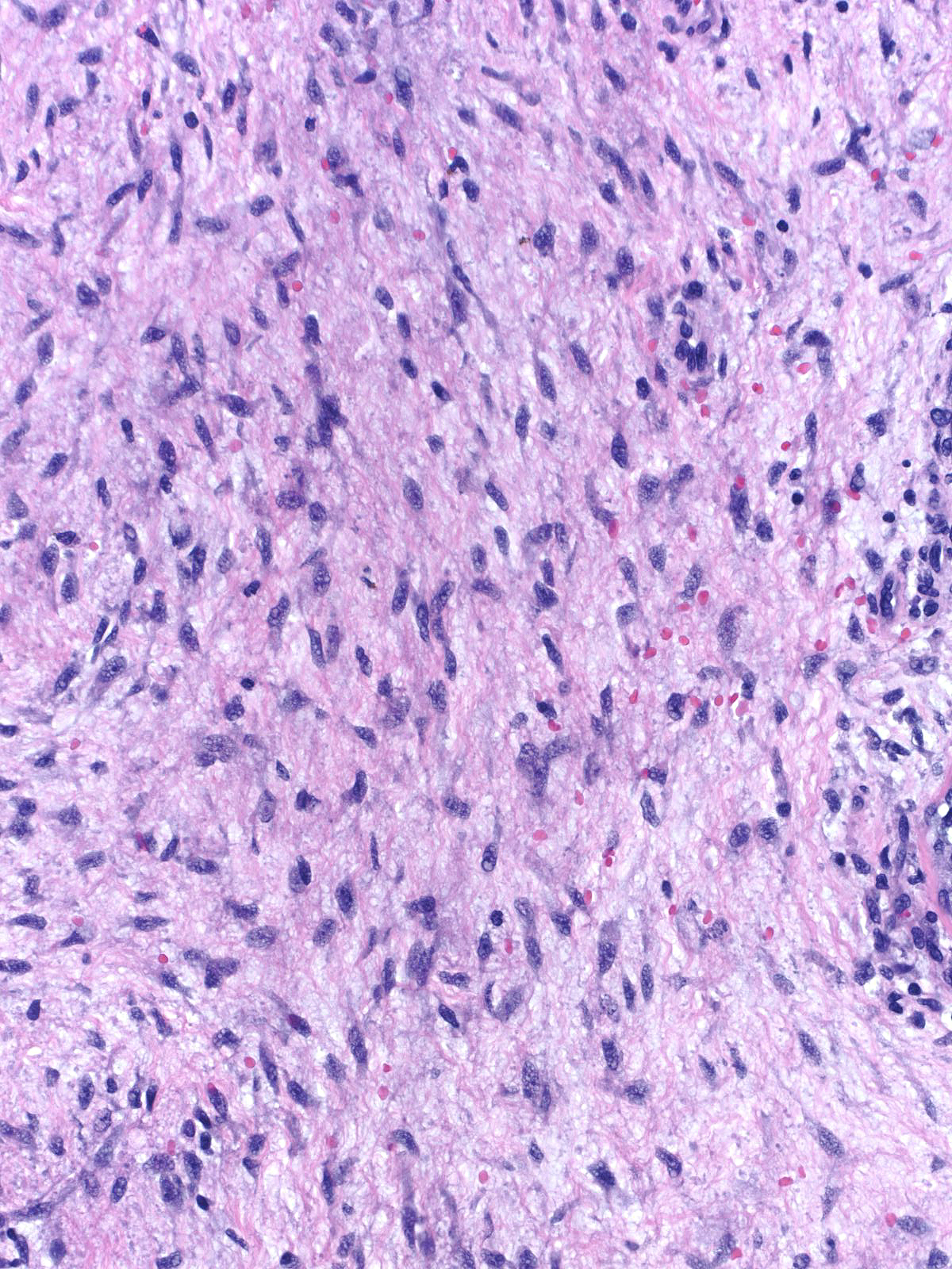
The cells possess atypical nuclei and scant cytoplasm. Depending of the grade of the tumor, the atypia can appear from minimal to marked.
 |
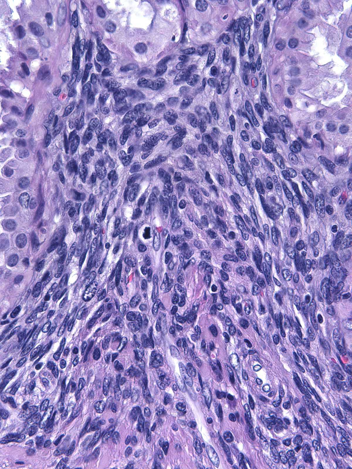 |
The stromal cells commonly display features of myofibroblasts and give rise to a pseudoangiomatous arrangement.
 |
 |
| Besides demonstrating characteristics of fibroblasts/myofibroblasts, the neoplastic cells can also take on morphological features of adipocytes, osteoblasts, chondroblasts, endothelial cells, or any other type of mesenchymal cell. In the presence of these forms of differentiation, the diagnosis remains phyllodes tumor rather than liposarcoma, osteosarcoma, chondrosarcoma, angiosarcoma, etc. (Neoplastic fat in a phyllodes tumor. Can you spot the lipoblasts?) | 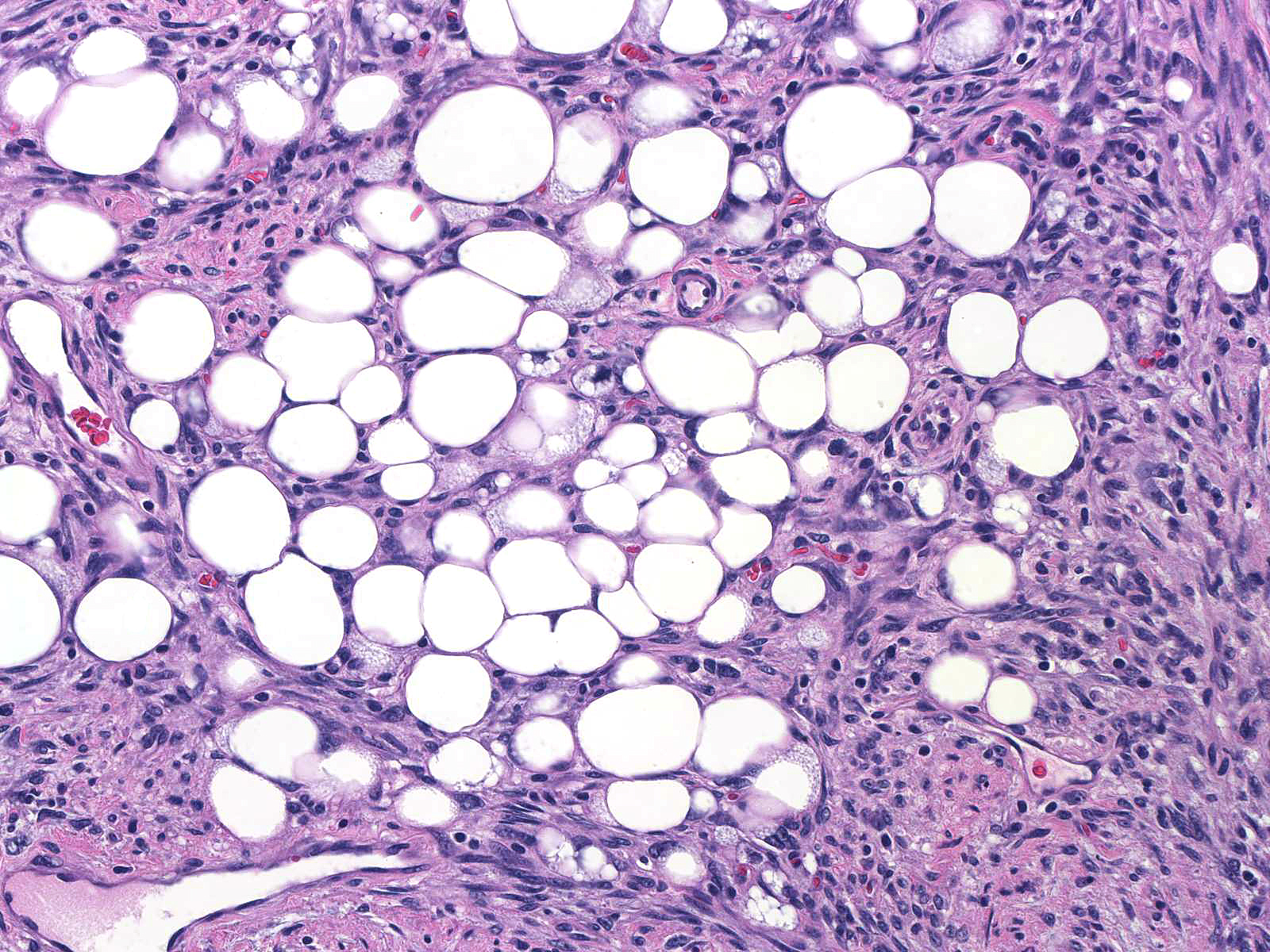 |
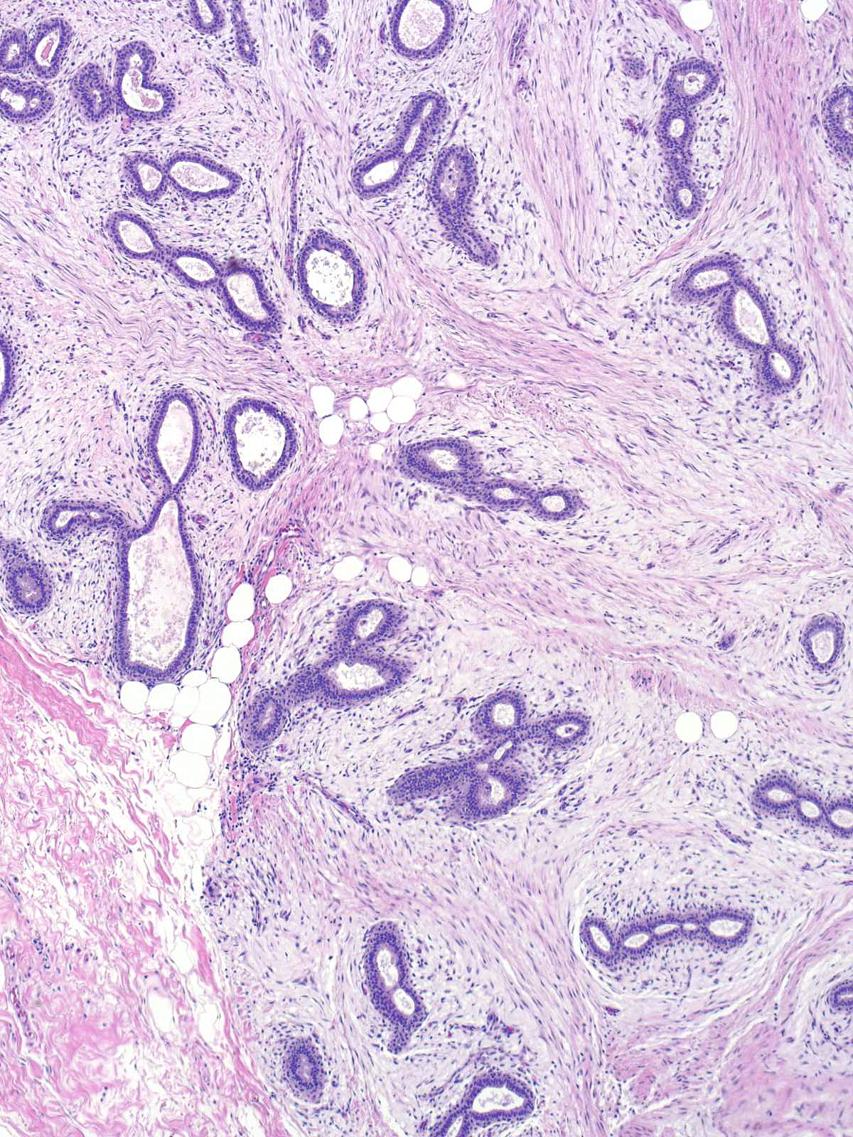
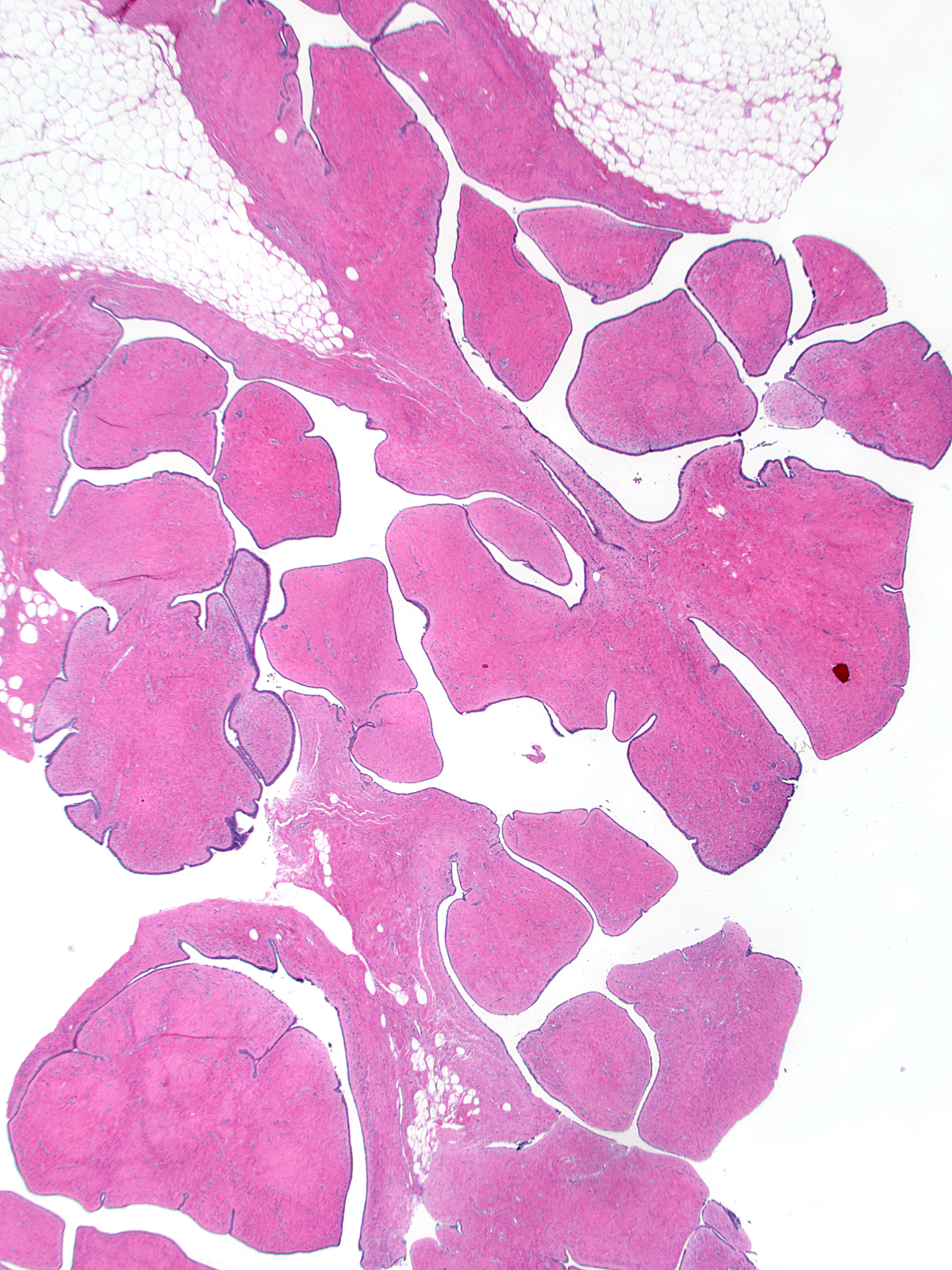
The extracellular matrix of phyllodes tumors consists of intermixed collagen and myxoid ground substances.
 |
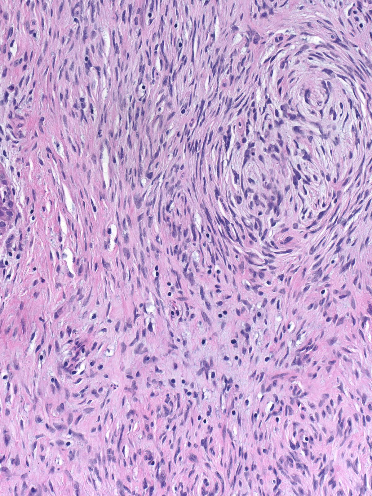 |
In rare tumors, one type of matrix can predominate.
 |
 |
| In other tumors, the predominant type of matrix can vary from region to region. | 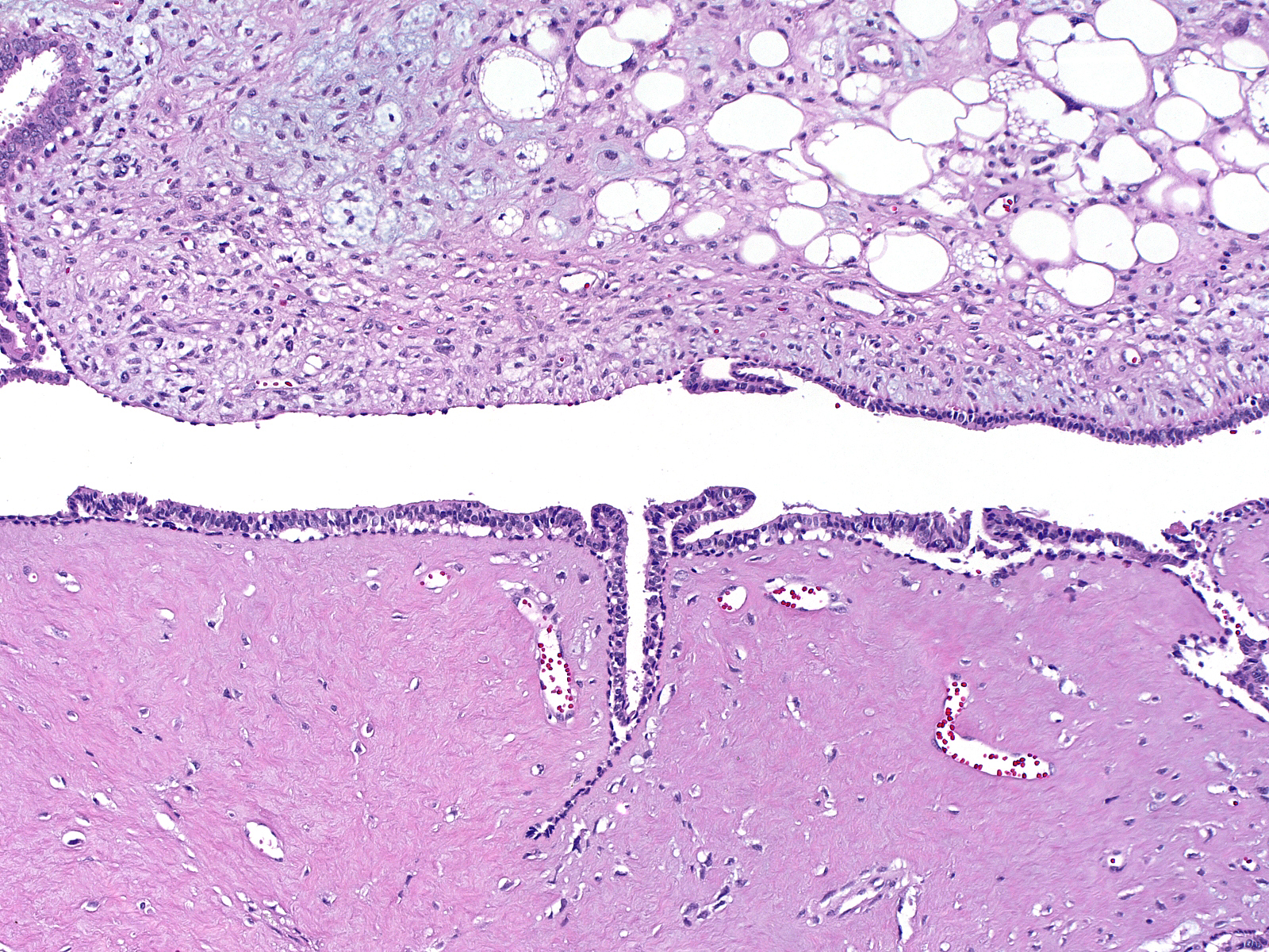 |
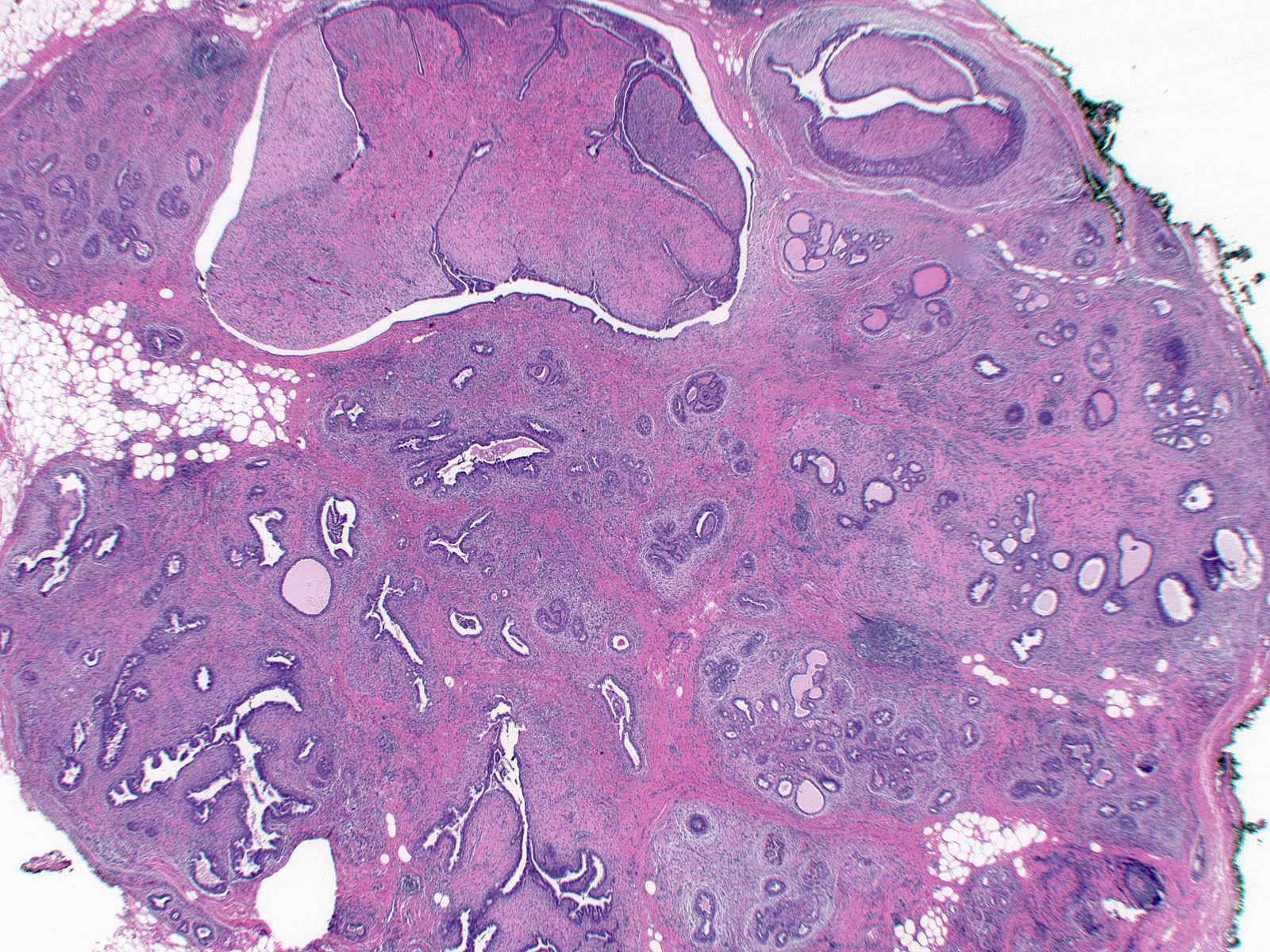
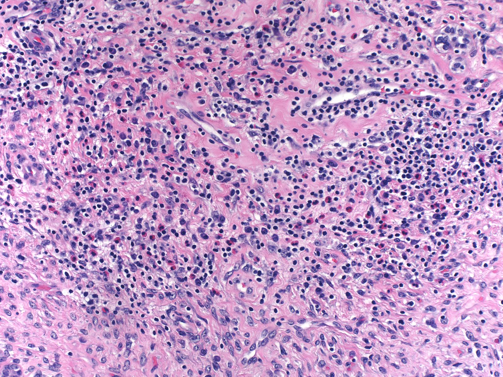
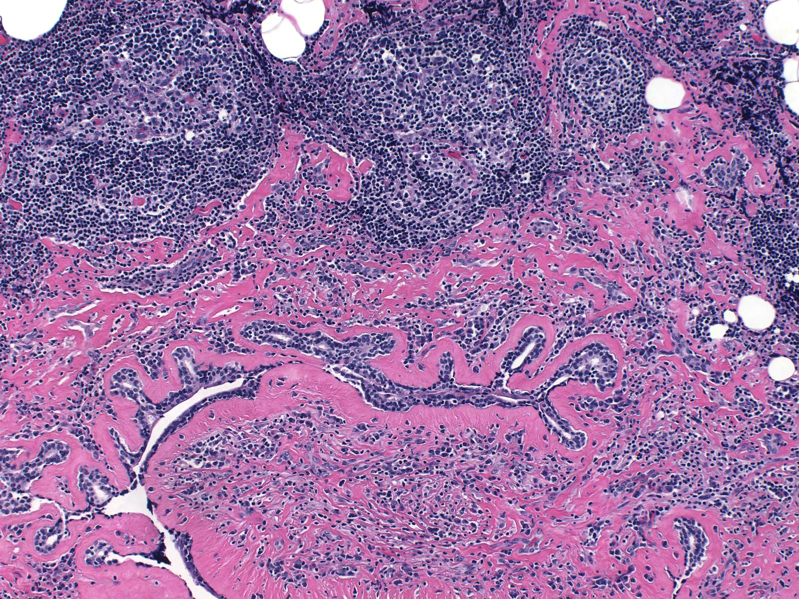
The stroma typically contains a few chronic inflammatory cells, but they can be present in great abundance. This example displays an eye-catching number of eosinophils, plasma cells, and lymphocytes, and the latter form two germinal centers at the edge of the mass.
 |
 |
 |
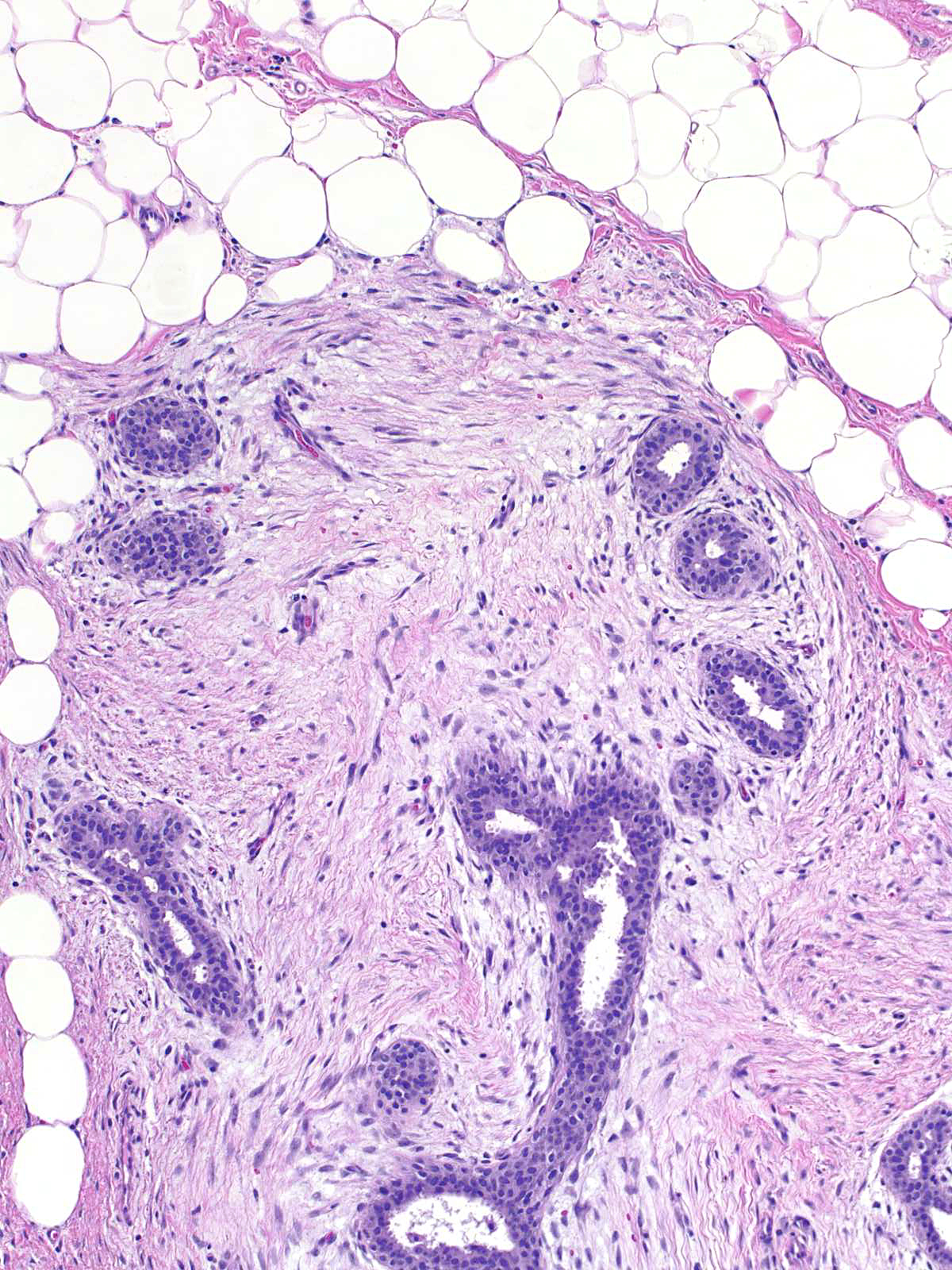
Many phyllodes tumors demonstrate invasion of the surrounding tissue by the neoplastic cells. The invasion is often subtle, but it can be obvious.
 |
 |
| The epithelium of the entrapped glands usually demonstrates evidence of proliferation such as cellular and nuclear enlargement, mitotic activity of the epithelial cells, and prominence of the myoepithelial cells. | 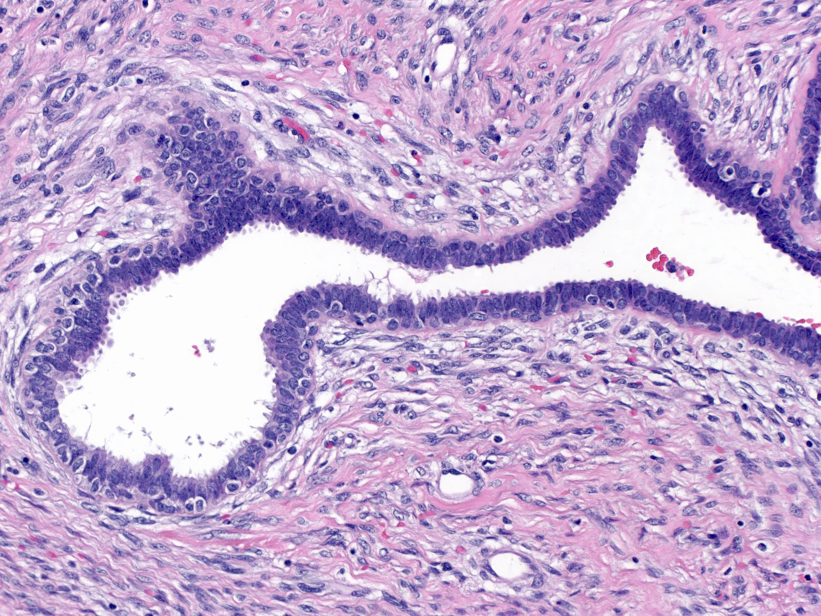 |
| Compare the attenuated epithelium of the fibroadenoma on the left with the proliferative epithelium of the phyllodes tumor on the right. |  |
| Growth of the epithelium in a flat pattern causes the glands to dilate and to assume unusual shapes likened to those of animals. | 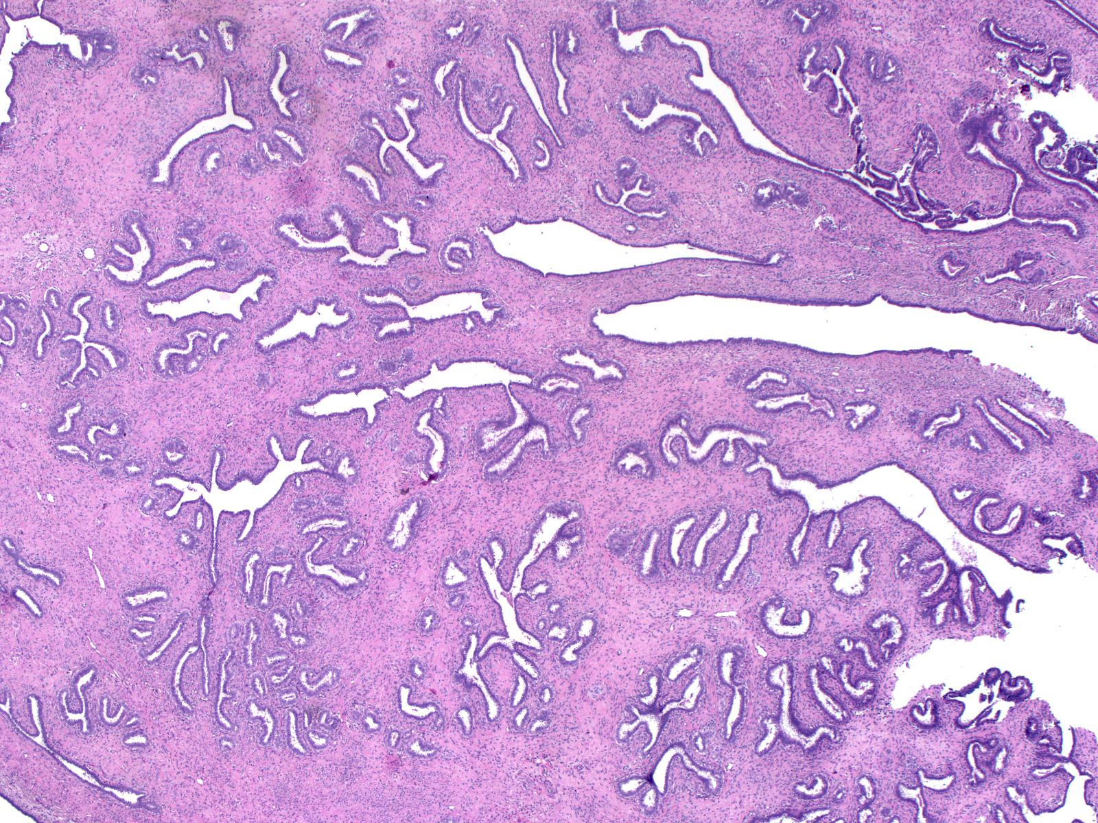 |
| The epithelium can give rise to ductal hyperplasia, which can appear especially florid. | 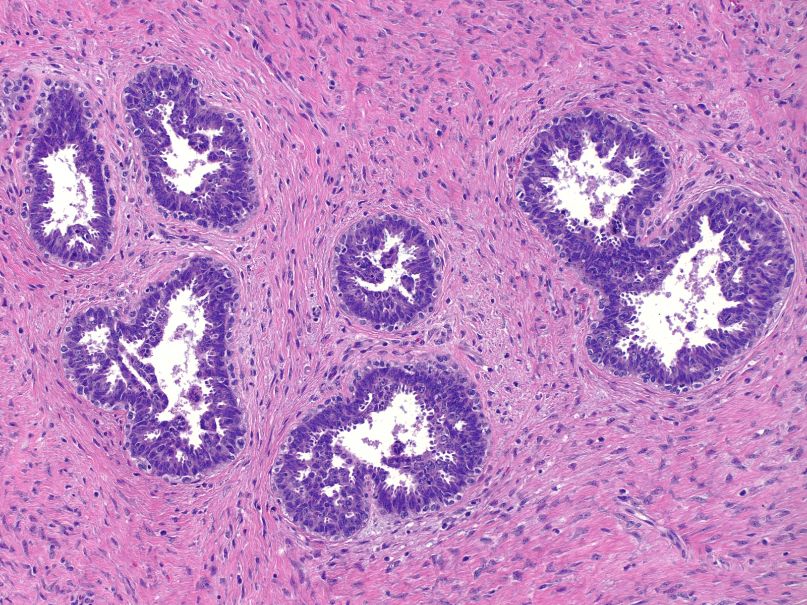 |
| In classic phyllodes tumors, the stroma forms fronds projecting into the dilated epithelial spaces. | 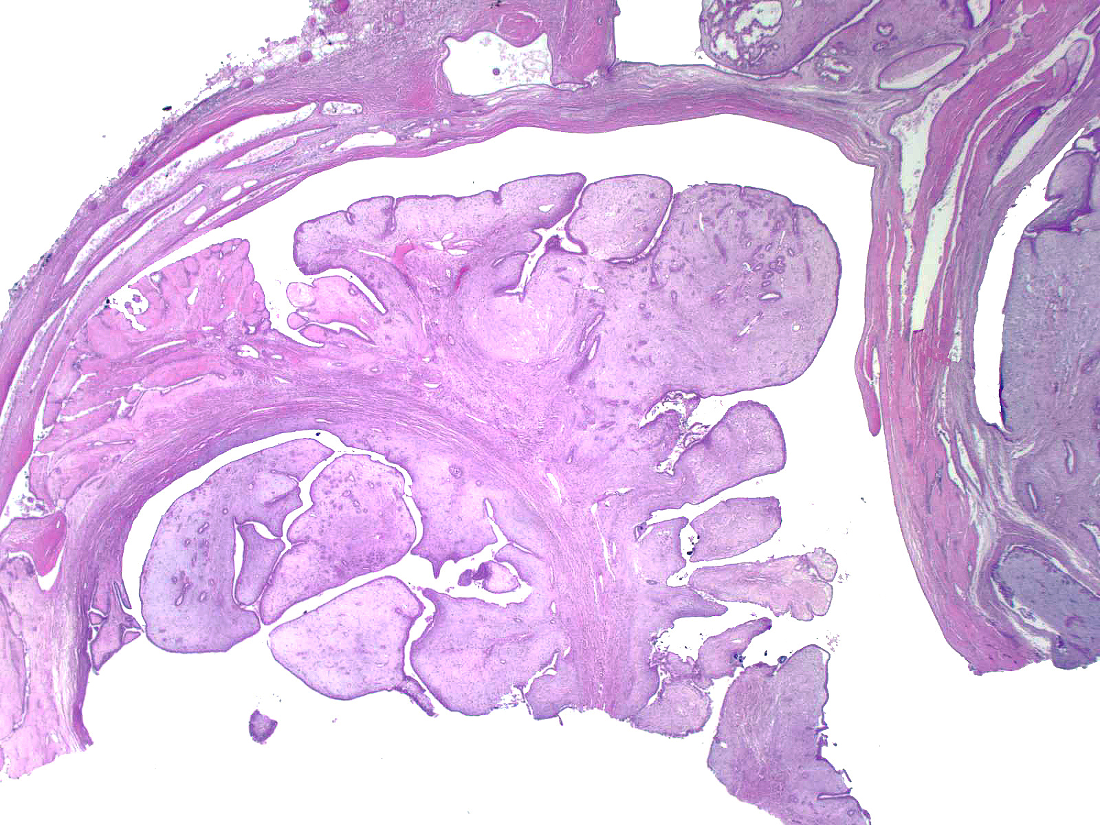 |
| The presence of fronds strongly suggests the diagnosis of phyllodes tumor, but many examples such as the one shown on the right do not form these structures. | 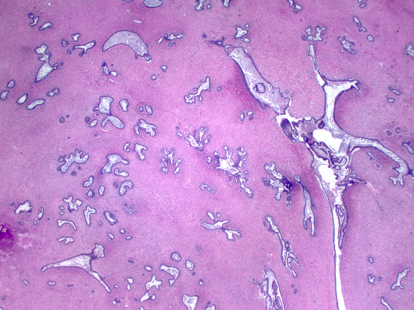 |
| The fronds form because dilatation of the entrapped glands creates spaces into which tongues of stromal cells protrude. | 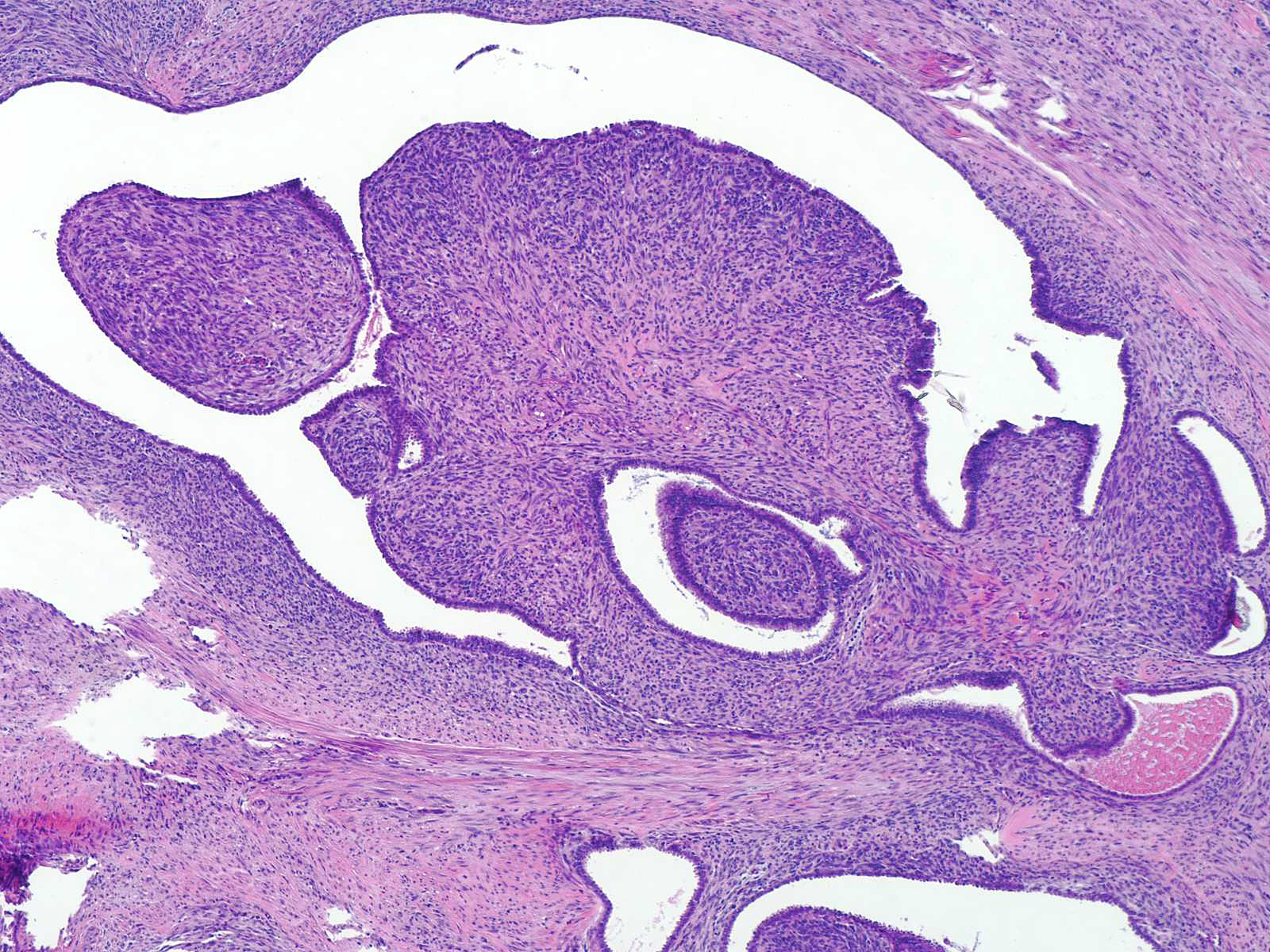 |
Pathologists grade phyllodes tumor using characteristics of the stromal cells. Low-grade tumors, also called benign phyllodes tumor, demonstrate minimal atypia, modest cellularity, and a low mitotic rate. High-grade tumors, also termed malignant phyllodes tumor, display marked cellularity, marked atypia, and a high mitotic rate. Tumors showing features between these extremes represent intermediate-grade phyllodes tumors. Foci of heterologous differentiation, hemorrhage, and necrosis would customarily indicate a high-grade phyllodes tumor.
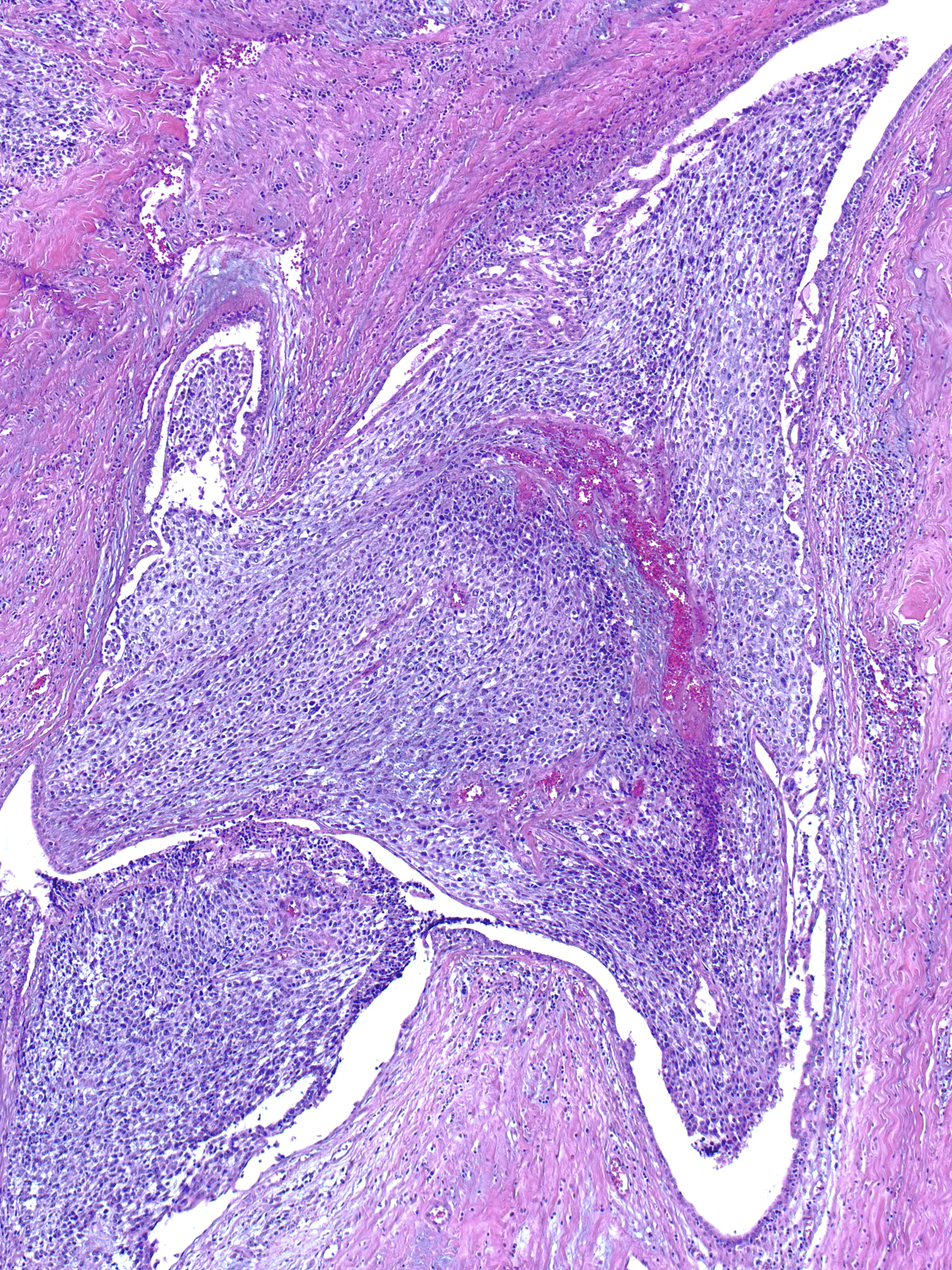 |
 |
Stromal overgrowth consists of a 40x field (4x objective and 10x ocular) composed solely of stroma. This phenomenon does not occur in most phyllodes tumors, and the diagnosis of phyllodes tumor does not depend on its presence. Stromal overgrowth may indicate a heightened likelihood of metastasis.
Phyllodes tumors usually arise from pre-existing fibroepithelial tumors. Fibroadenomas probably represent the most common lesion of origin, but phyllodes tumors can also arise from hamartomas. The presence of such an underlying fibroepithelial tumor contributes to the regional variability in glandular architecture, stromal cellularity, and composition of the extracellular matrix that characterize phyllodes tumors.
 |
 |