Difference between revisions of "Myofibroblastoma"
(Created page with "__NOCACHE__{{DISPLAYTITLE:Myofibroblastoma}} {{:TOC}} == General == Image:29-1 10_x_A.jpg|thumb|Myofibroblastomas are benign tumors composed of bland myofibroblasts interspe...") |
|||
| (3 intermediate revisions by 2 users not shown) | |||
| Line 1: | Line 1: | ||
| − | __NOCACHE__ | + | __NOCACHE__ |
{{:TOC}} | {{:TOC}} | ||
| − | == | + | <br> |
| − | + | == Introduction == | |
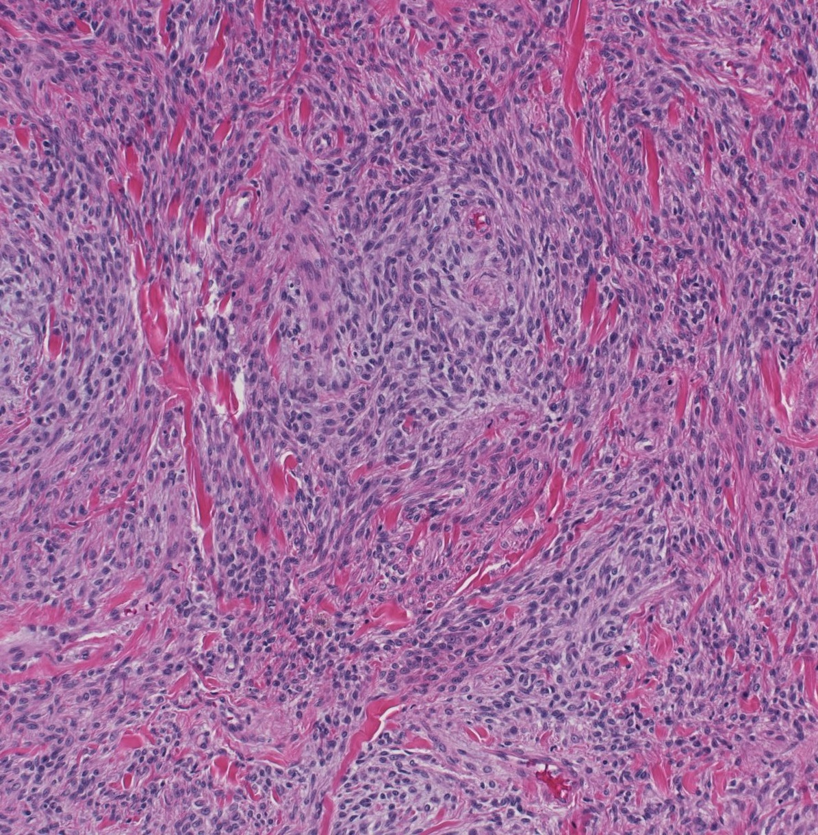
| − | + | {{dxintronodisc|Myofibroblastoma|A myofibroblastoma is a tumor composed of benign myofibroblasts.|Patients with myofibroblastomas are usually middle aged or older. Early reports suggested that the tumor occurs mostly in men, but later experience indicates that the lesion arises in men and women with approximately equal frequency. The tumors usually present as a mass evident on palpation or radiological imaging.|Myofibroblastomas grow as well-defined, bulging nodules of homogeneous, grey-to-pink tissue. They do not display hemorrhage or cystic degeneration.|Mammary myofibroblastomas look like corresponding tumors arising in other organs. Typical mammary myofibroblastomas feature bland spindle cells interspersed among thick bundles of collagen.|Nodular pseudoangiomatous stromal hyperplasia (glandular component), spindle cell carcinoma (cytological atypia), sarcoma (cytological atypia), fibromatosis (sweeping fascicles, infiltrative border, expression of ß-catenin)||29-1 10_x_A.jpg|Myofibroblastomas are benign tumors composed of bland myofibroblasts interspersed among thick bands of collagen.}} | |
| − | |||
| − | |||
| − | |||
| − | |||
| − | |||
| − | |||
| − | |||
| − | |||
| − | |||
{{img1|Classic myofibroblastoma has a well defined border and compresses the surrounding parenchyma. In a minority of cases, the neoplastic cells infiltrate the adjacent tissue. The mass does not harbor glandular tissue within it.|29-2 Low_power-3.jpg}} | {{img1|Classic myofibroblastoma has a well defined border and compresses the surrounding parenchyma. In a minority of cases, the neoplastic cells infiltrate the adjacent tissue. The mass does not harbor glandular tissue within it.|29-2 Low_power-3.jpg}} | ||
{{img1|Bundles of uniform, slender, bipolar spindle cells arranged in short fascicles and interspersed broad bands of hyalinized collagen comprise the mass. The collagen exhibits a dense, eosinophilic hue and a homogenous appearance. One occasionally finds a perivascular infiltrate of chronic inflammatory cells.|29-3 Cells-6.jpg}} | {{img1|Bundles of uniform, slender, bipolar spindle cells arranged in short fascicles and interspersed broad bands of hyalinized collagen comprise the mass. The collagen exhibits a dense, eosinophilic hue and a homogenous appearance. One occasionally finds a perivascular infiltrate of chronic inflammatory cells.|29-3 Cells-6.jpg}} | ||
| Line 20: | Line 11: | ||
{{img1|Tumors with extensive adipocytic differentiation have been called lipomatous myofibroblastomas. These cases hint at relationship between myofibroblastomas and spindle cell lipomas.|29-6 Lipomatous.jpg}} | {{img1|Tumors with extensive adipocytic differentiation have been called lipomatous myofibroblastomas. These cases hint at relationship between myofibroblastomas and spindle cell lipomas.|29-6 Lipomatous.jpg}} | ||
{{img1|The neoplastic cells can also assume an epithelioid shape; one refers to such examples as epithelioid myofibroblastomas. This type of myofibroblastoma closely mimics an invasive lobular carcinoma. Attention to the dishesive properties of the neoplastic cells, their cytological atypia, and their expression of keratin will distinguish invasive lobular carcinoma from myofibroblastoma.|29-5 Epithelioid.jpg}} | {{img1|The neoplastic cells can also assume an epithelioid shape; one refers to such examples as epithelioid myofibroblastomas. This type of myofibroblastoma closely mimics an invasive lobular carcinoma. Attention to the dishesive properties of the neoplastic cells, their cytological atypia, and their expression of keratin will distinguish invasive lobular carcinoma from myofibroblastoma.|29-5 Epithelioid.jpg}} | ||
| − | + | {{:mgh:breast-footer}} | |
| − | |||
| − | |||
Latest revision as of 08:41, July 17, 2020
Contents
Introduction
|
Definition: A myofibroblastoma is a tumor composed of benign myofibroblasts. Clinical Significance: Patients with myofibroblastomas are usually middle aged or older. Early reports suggested that the tumor occurs mostly in men, but later experience indicates that the lesion arises in men and women with approximately equal frequency. The tumors usually present as a mass evident on palpation or radiological imaging. Gross Findings: Myofibroblastomas grow as well-defined, bulging nodules of homogeneous, grey-to-pink tissue. They do not display hemorrhage or cystic degeneration. Microscopic Findings: Mammary myofibroblastomas look like corresponding tumors arising in other organs. Typical mammary myofibroblastomas feature bland spindle cells interspersed among thick bundles of collagen. Differential Diagnosis: Nodular pseudoangiomatous stromal hyperplasia (glandular component), spindle cell carcinoma (cytological atypia), sarcoma (cytological atypia), fibromatosis (sweeping fascicles, infiltrative border, expression of ß-catenin) |
 |
| Classic myofibroblastoma has a well defined border and compresses the surrounding parenchyma. In a minority of cases, the neoplastic cells infiltrate the adjacent tissue. The mass does not harbor glandular tissue within it. | 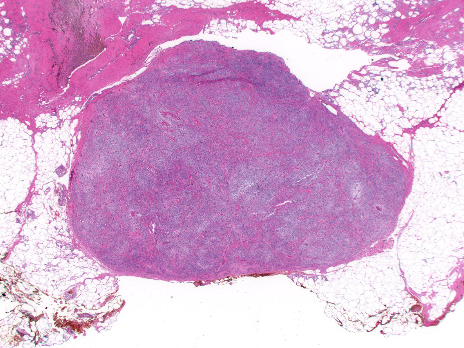 |
| Bundles of uniform, slender, bipolar spindle cells arranged in short fascicles and interspersed broad bands of hyalinized collagen comprise the mass. The collagen exhibits a dense, eosinophilic hue and a homogenous appearance. One occasionally finds a perivascular infiltrate of chronic inflammatory cells. | 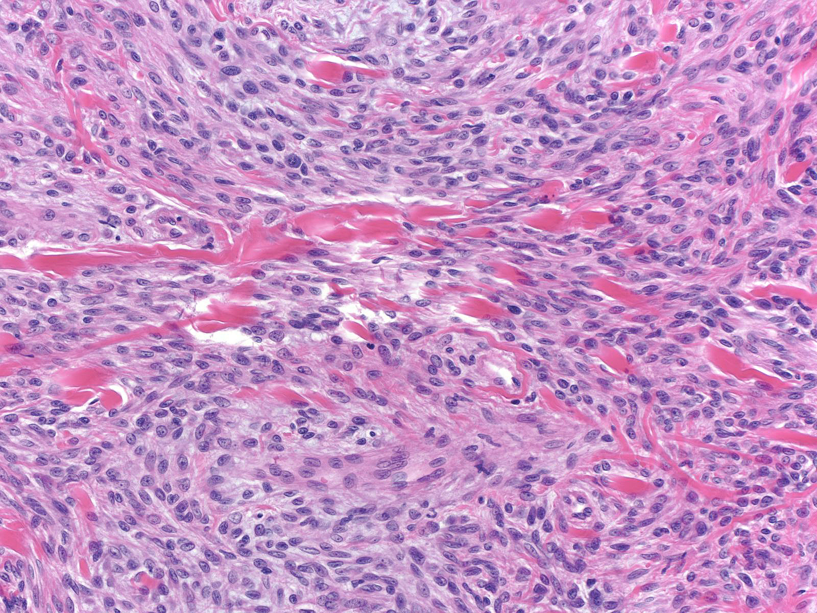 |
| The spindle cells have bland, ovoid to spindled nuclei with dispersed chromatin and small nucleoli. Nuclear grooves may be present. The cytoplasm appears pale and eosinophilic, and the cell borders look indistinct. One finds mitotic figures and multinucleate cells only rarely, but cellular degeneration can give rise to isolated pleomorphic cells. | 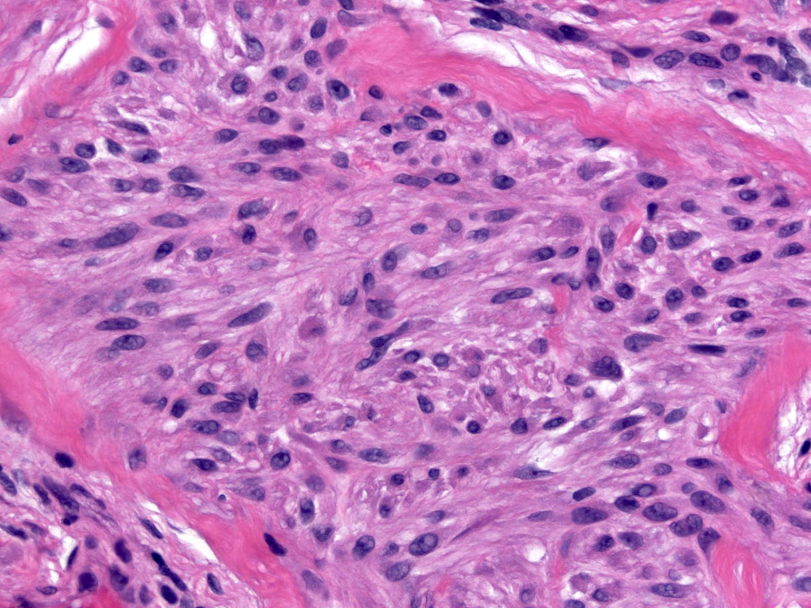 |
Myofibroblastomas can demonstrate many variations from the classic pattern. The spindle cells can differentiate to form fat, smooth muscle, or cartilage. In cellular myofibroblastomas, the spindle cells dominate, whereas collagenized myofibroblastomas consist mostly of dense collagen. Myxoid myofibroblastomas feature myxoid extracellular material rather than collagen.
| Tumors with extensive adipocytic differentiation have been called lipomatous myofibroblastomas. These cases hint at relationship between myofibroblastomas and spindle cell lipomas. |  |
| The neoplastic cells can also assume an epithelioid shape; one refers to such examples as epithelioid myofibroblastomas. This type of myofibroblastoma closely mimics an invasive lobular carcinoma. Attention to the dishesive properties of the neoplastic cells, their cytological atypia, and their expression of keratin will distinguish invasive lobular carcinoma from myofibroblastoma. | 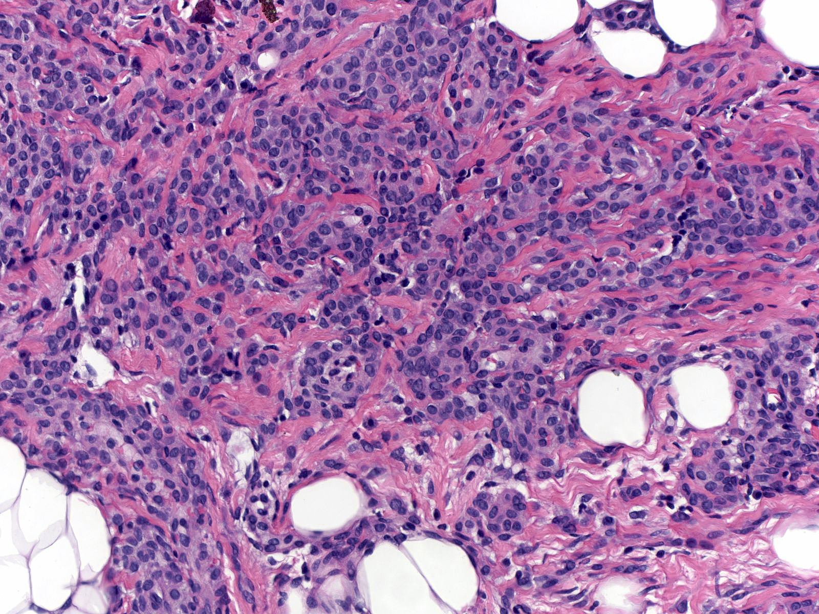 |