Difference between revisions of "mgh:breast-Immunohistochemistry in Breast Pathology"
| (2 intermediate revisions by one other user not shown) | |||
| Line 25: | Line 25: | ||
{{img1|Hyperplastic ductal cells can sometimes stain for p63.|7-15 p63_UDH_20_x_M.jpg}} | {{img1|Hyperplastic ductal cells can sometimes stain for p63.|7-15 p63_UDH_20_x_M.jpg}} | ||
Scattered cells in uncommon examples of IDC and in common examples of papillary carcinoma stain for p63. | Scattered cells in uncommon examples of IDC and in common examples of papillary carcinoma stain for p63. | ||
| − | {{ | + | {{img4|7-16 IDC_10_x.jpg|7-17 p63_IDC_10_x.jpg|7-18 p63_Pap_CA_20_x.jpg|Invasive ductal carcinoma (H&E)|Invasive ductal carcinoma (p63)|Papillary carcinoma (p63)}} |
Neoplastic myoepithel cells and the cells of spindle cell carcinoma regularly stain for p63. | Neoplastic myoepithel cells and the cells of spindle cell carcinoma regularly stain for p63. | ||
| − | {{ | + | {{img4|7-19 p63_Adenosq_20_x.jpg|7-20 Spindle_cell_20_x_M_p63.jpg|Adenosquamous carcinoma|Spindle cell carcinoma}} |
== Smooth Muscle Myosin Heavy Chain == | == Smooth Muscle Myosin Heavy Chain == | ||
Several types of cells other than myoepithelial cells express this protein. Pay attention to the features evident on the H&E-stained sections to avoid misinterpretations. | Several types of cells other than myoepithelial cells express this protein. Pay attention to the features evident on the H&E-stained sections to avoid misinterpretations. | ||
| − | {{ | + | {{img4|7-21 Myosin_Endothelial_20_x.jpg|7-22 Myosin_Lymphoid_40_x.jpg|Endothelial cells Lymphoid cells (above)|myoepithelial cells (below)}} |
== Calponin == | == Calponin == | ||
Antibodies to calponin frequently react with stroma myofibroblasts. When the myofibroblasts sit next to epithelial cells, one could mistake the stromal cells for myoepithelial cells and thereby fail to recognize the invasive nature of carcinoma cells. | Antibodies to calponin frequently react with stroma myofibroblasts. When the myofibroblasts sit next to epithelial cells, one could mistake the stromal cells for myoepithelial cells and thereby fail to recognize the invasive nature of carcinoma cells. | ||
| Line 44: | Line 44: | ||
Do not confuse the granular staining of myoepithelial cells with the linear staining of lymphatic endothelial cells. | Do not confuse the granular staining of myoepithelial cells with the linear staining of lymphatic endothelial cells. | ||
{{img4|7-27 D2-40_Mye_40_x.jpg|7-28 D2-40_LVI_40_x.jpg|Myoepithelial cells|Endothelial cells}} | {{img4|7-27 D2-40_Mye_40_x.jpg|7-28 D2-40_LVI_40_x.jpg|Myoepithelial cells|Endothelial cells}} | ||
| + | {{:mgh:breast-footer}} | ||
Latest revision as of 18:08, July 13, 2020
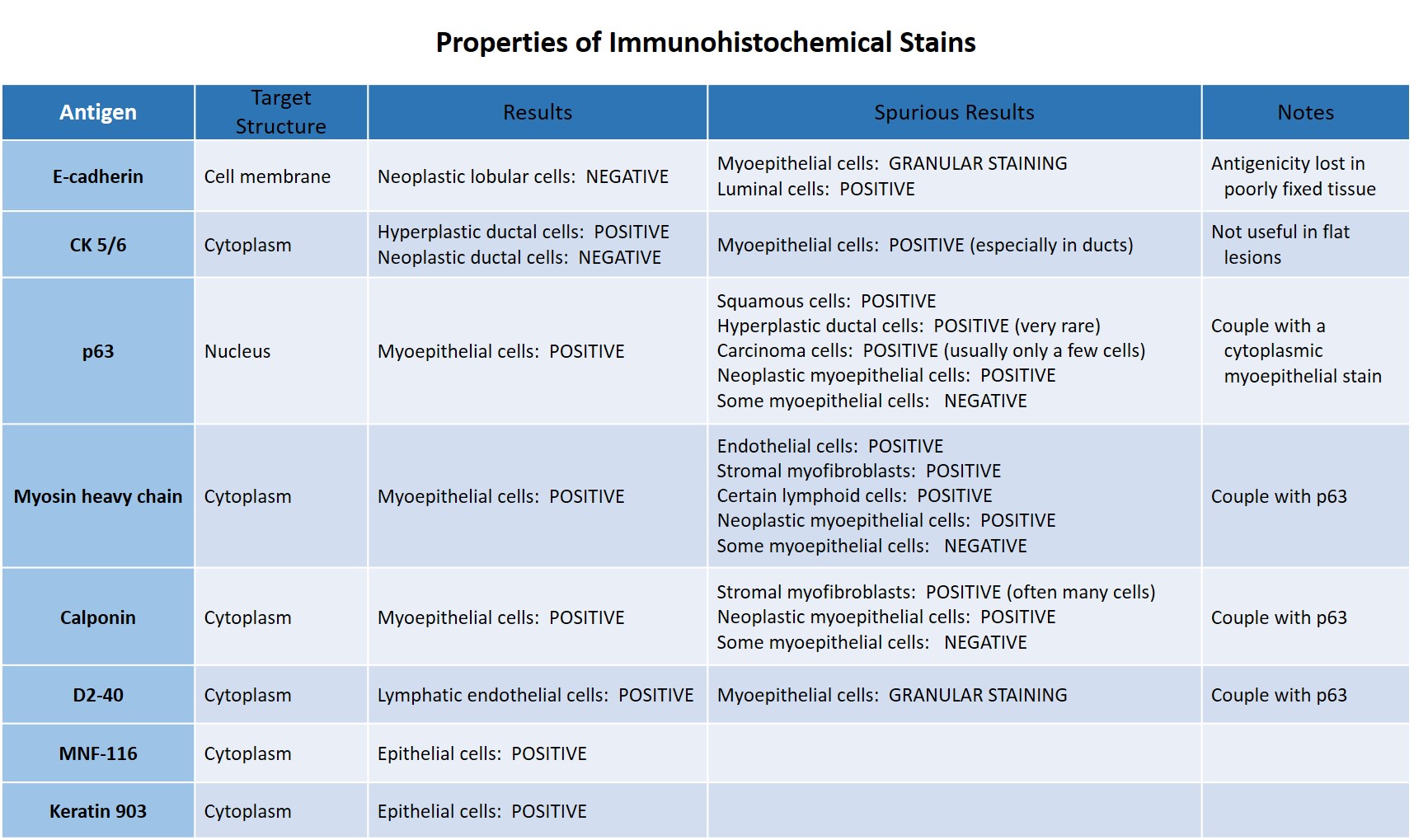
The following table lists several commonly used stains in the study of breast pathology, their expected results, and several causes of spurious staining.
 |
|
Here are a few hints regarding interpretation of immunohistochemical staining results:
E-cadherin
| Staining of both luminal cells and myoepithelial cells can make it difficult to appreciate the absence of staining of individual neoplastic lobular cells and of clusters of just two or three lobular cells. Look for the absence of membrane staining between adjacent neoplastic cells rather than between neoplastic cells and normal cells | 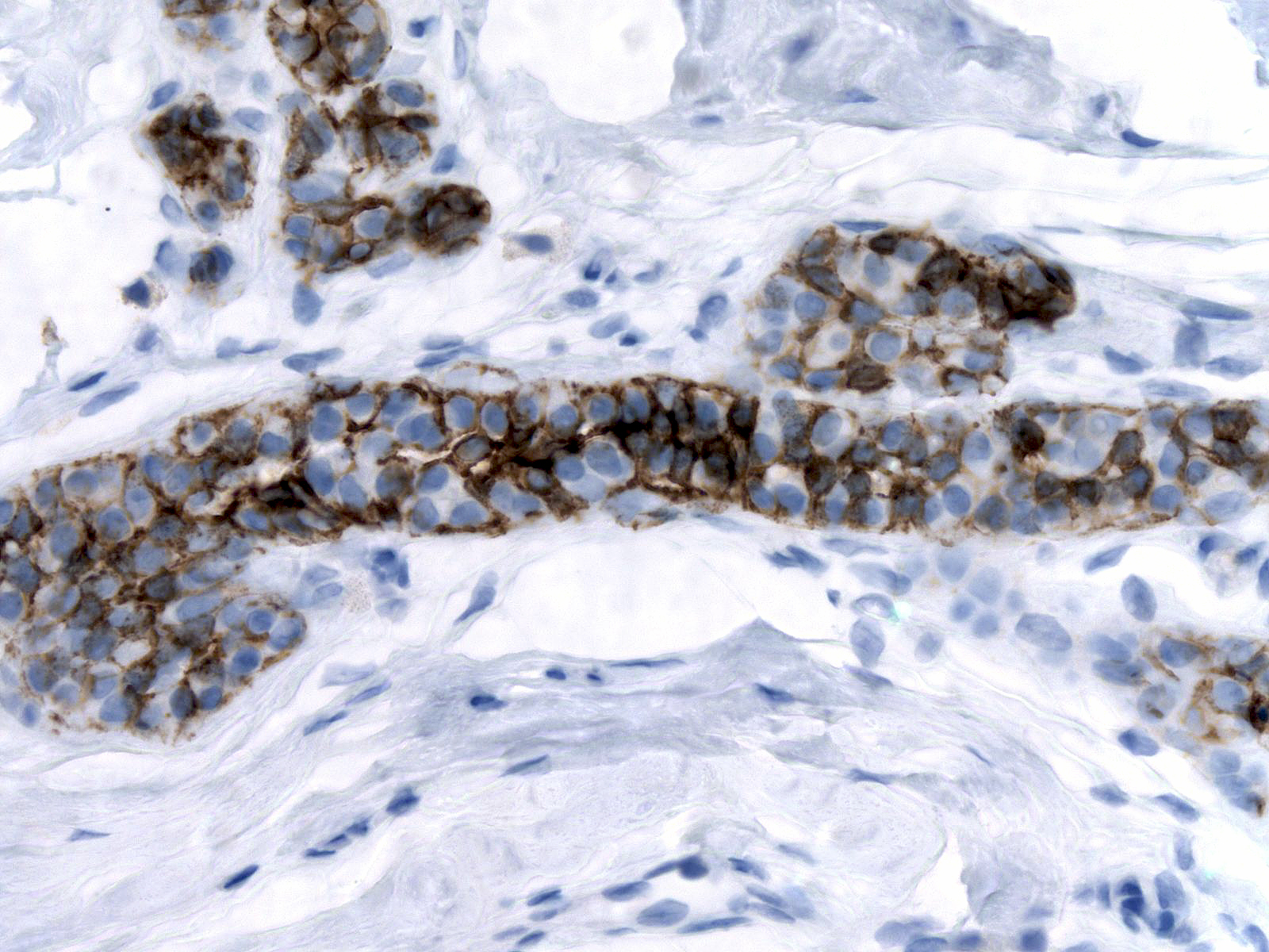 |
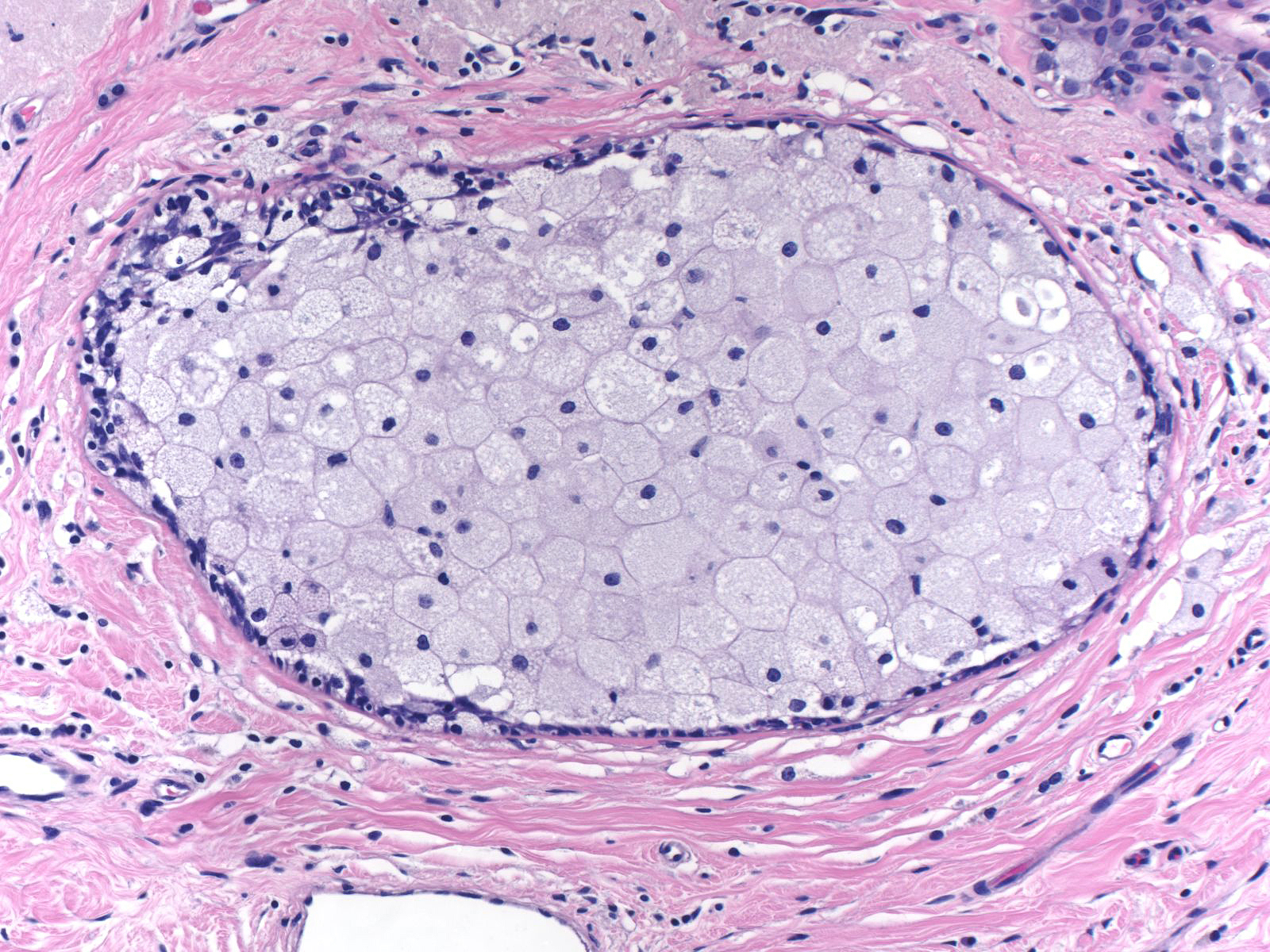
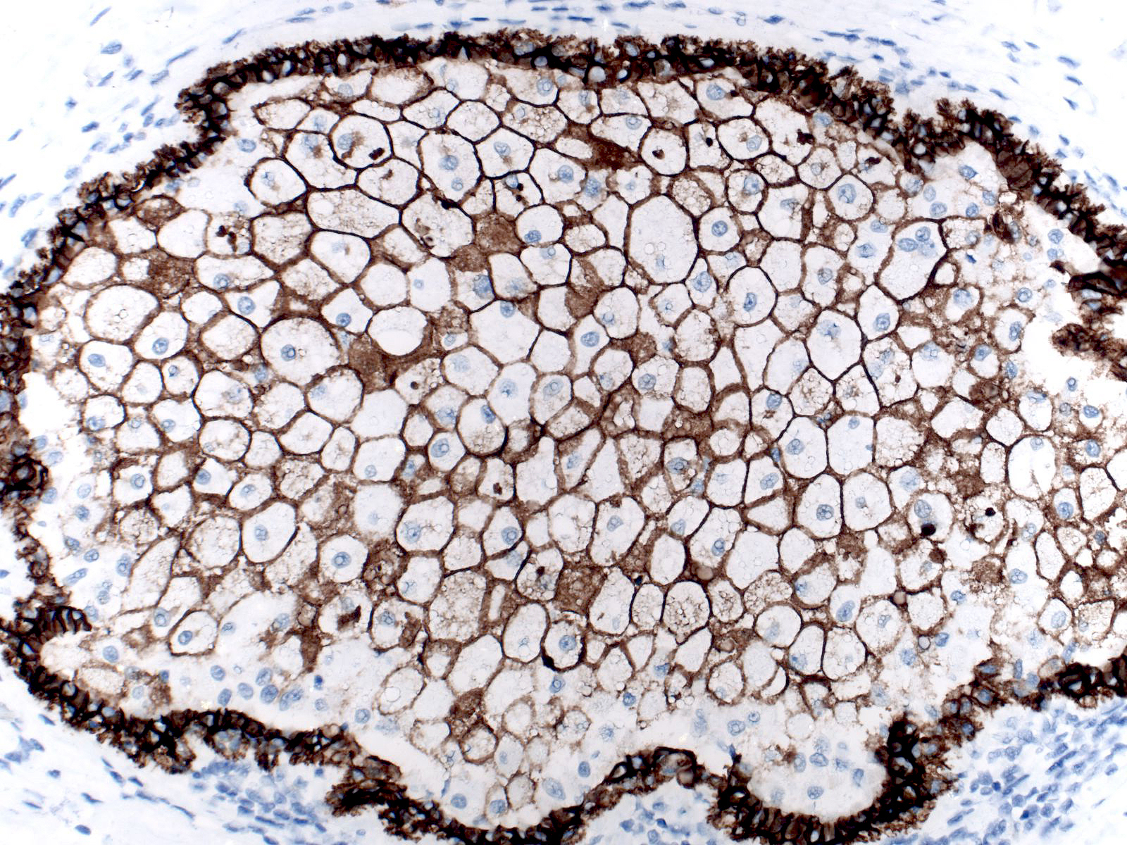
Under certain circumstances, histiocytes can display granular membranous staining.
 |
 |
 |
CK 5/6
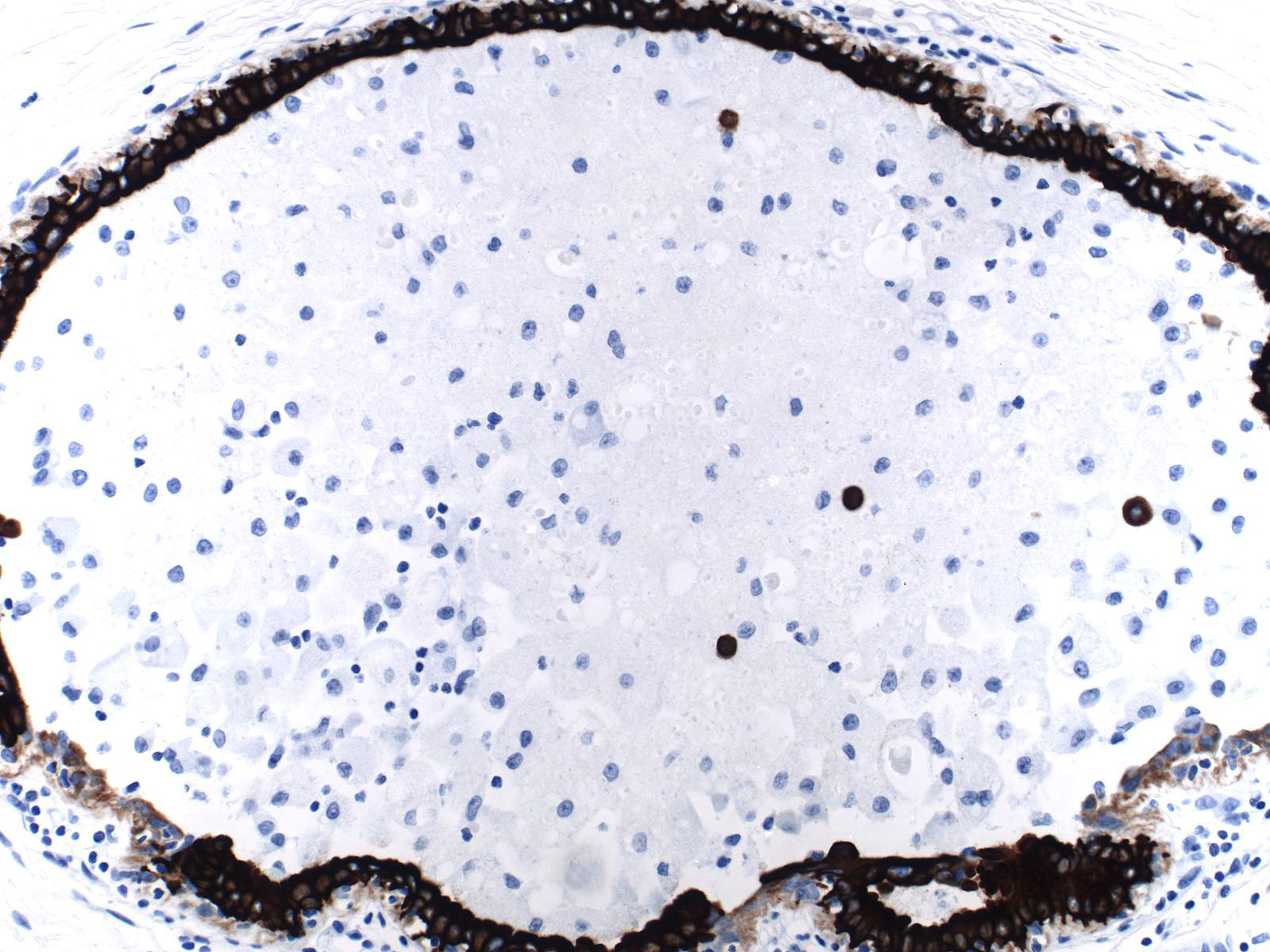
Most epithelial cells growing in a flat pattern fail to stain for CK 5/6; consequently, one cannot us staining for CK 5/6 to differentiate flat atypia from fibrocystic changes or flat ductal hyperplasia. Apocrine cells do not stain for CK 5/6, either. Integrating the morphological features evident on H&E-stained sections with the results of immunohistochemical staining will prevent you from confusing normal luminal cells, hyperplastic ductal cells, and apocrine cells with neoplastic ductal cells.
| The cytoplasm of these apocrine cells does not stain for CK 5/6, but the chromogen (DAB) reacts with the apical granules in a nonspecific way. | 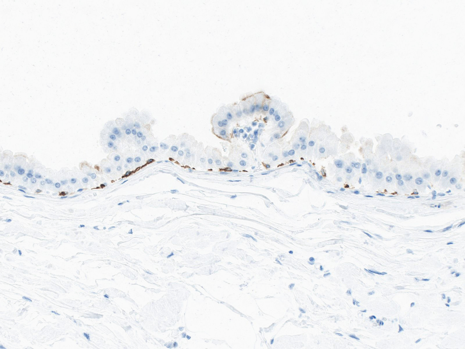 |
Some of the cells in this focus of UDH demonstrate apocrine change; consequently, they fail to stain for CK 5/6. By recognizing the apocrine nature of these cells, you will not mistake them for neoplastic cells and make an erroneous diagnosis such as ADH or DCIS.
 |
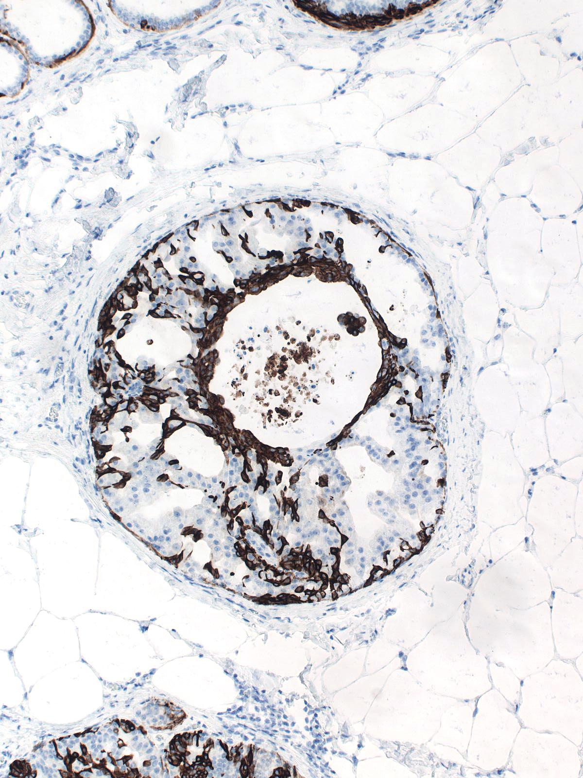 |
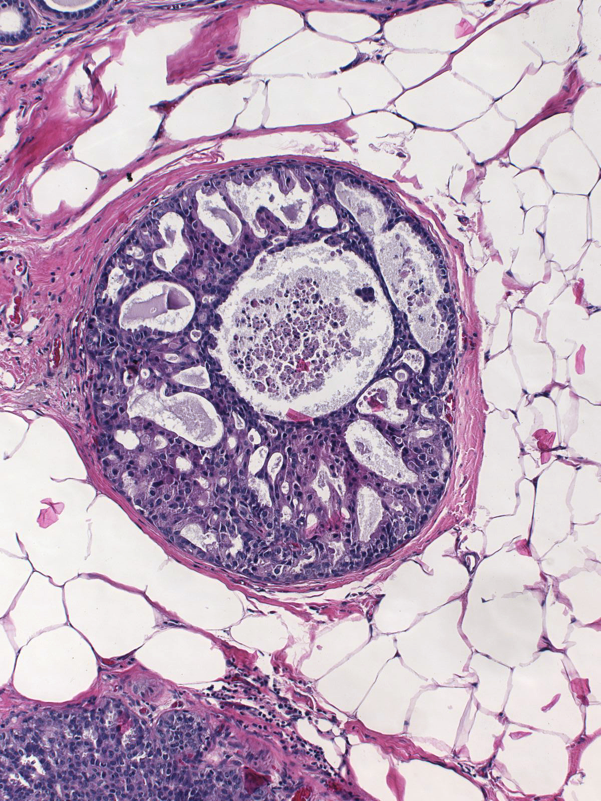
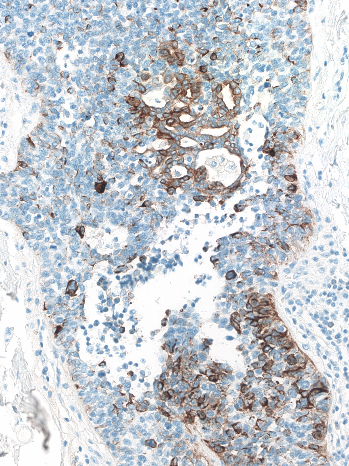
The expression of CK 5/6 does not guarantee the benign nature of an epithelial population. Uncommon intermediate‑ and high‑grade ductal carcinomas can stain for CK 5/6, and squamous carcinoma typically express this type of keratin.
 |
 |
|
This intermediate grade DCIS stains for CK 5/6 in a pattern that superficially resembles the pattern seen in UDH.
 |
 |
|
This high grade DCIS stains for CK 5/6 in a pattern that looks very much like the pattern seen in UDH.
 |
 |
|
This squamous carcinoma stains for CK 5/6 intensely.
p63
At times, myoepithelial cells fail to stain for p63, so the absence of this protein does not exclude the possibility that a cell is a myoepithelial cell. A positive result does not establish either the myoepithelial nature of a cell or the benign nature of a cell. As always, one must correlate the results of immunohistochemical staining with the findings evident on H&E-stained sections.
| Hyperplastic ductal cells can sometimes stain for p63. | 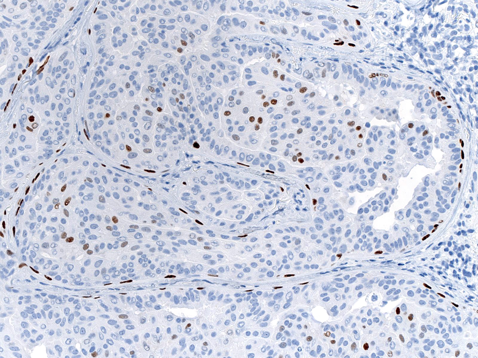 |
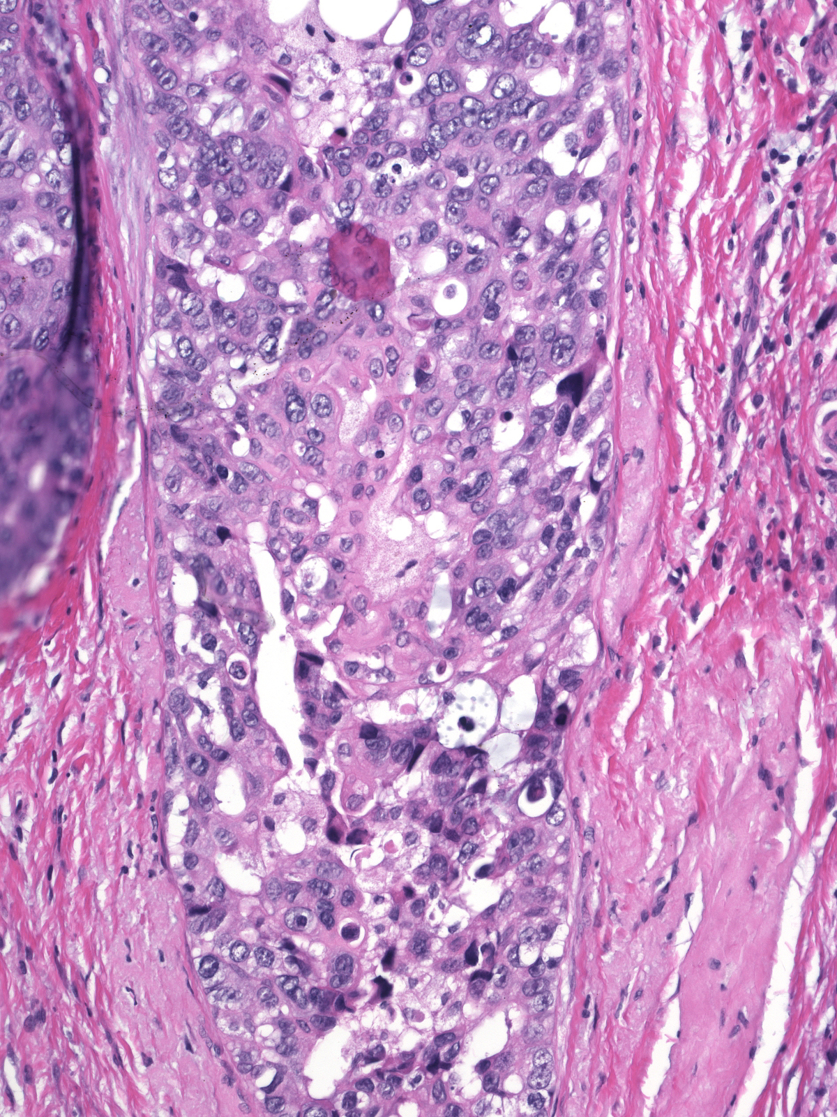
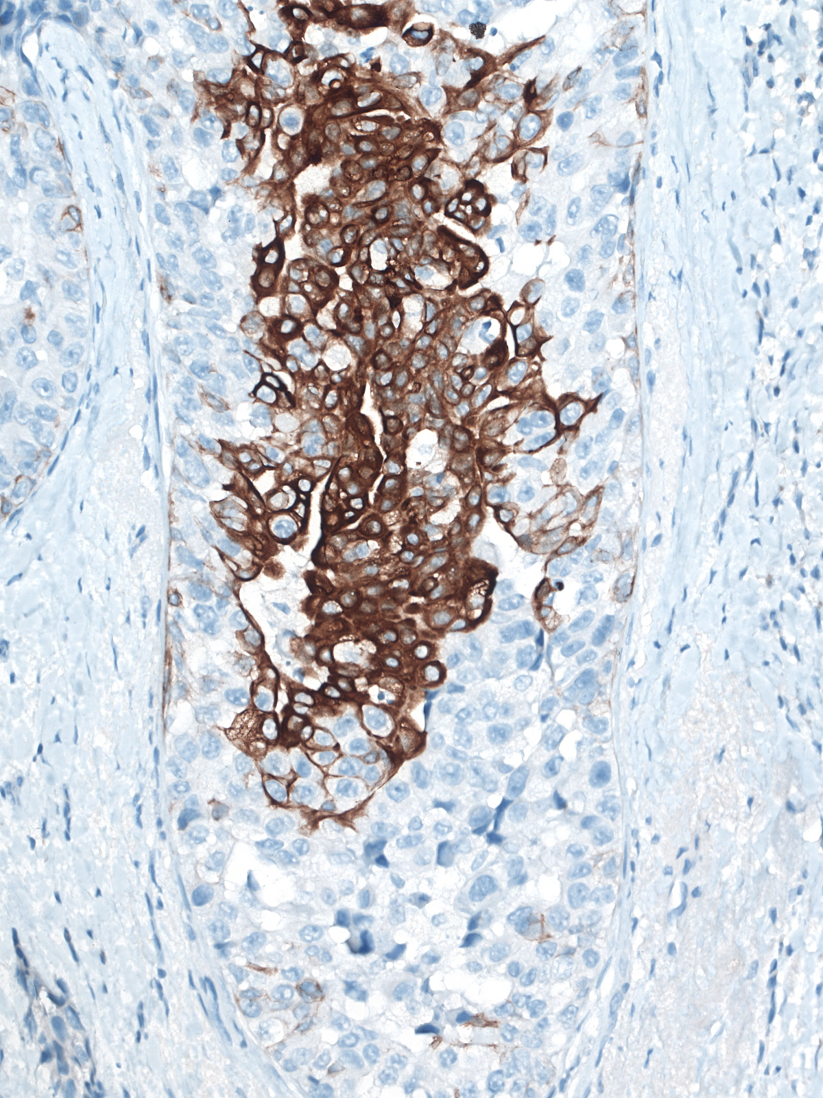
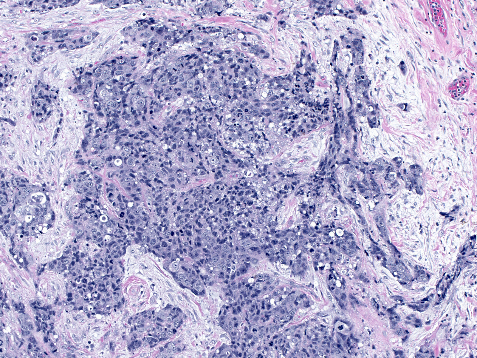
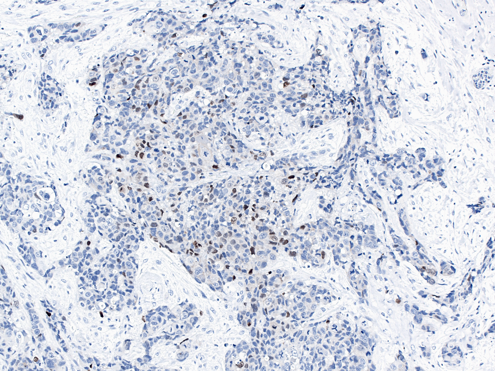
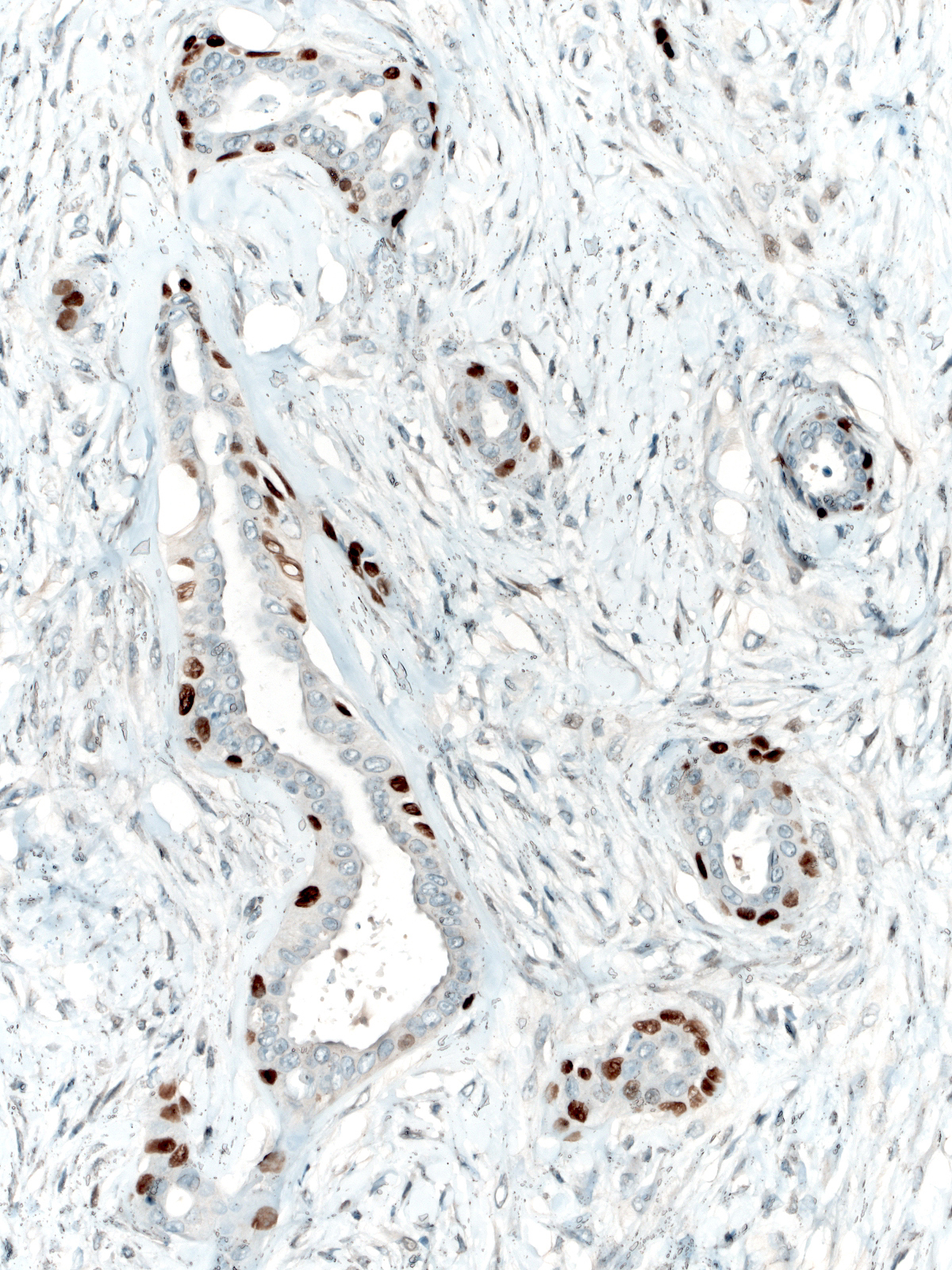
Scattered cells in uncommon examples of IDC and in common examples of papillary carcinoma stain for p63.
 |
 |
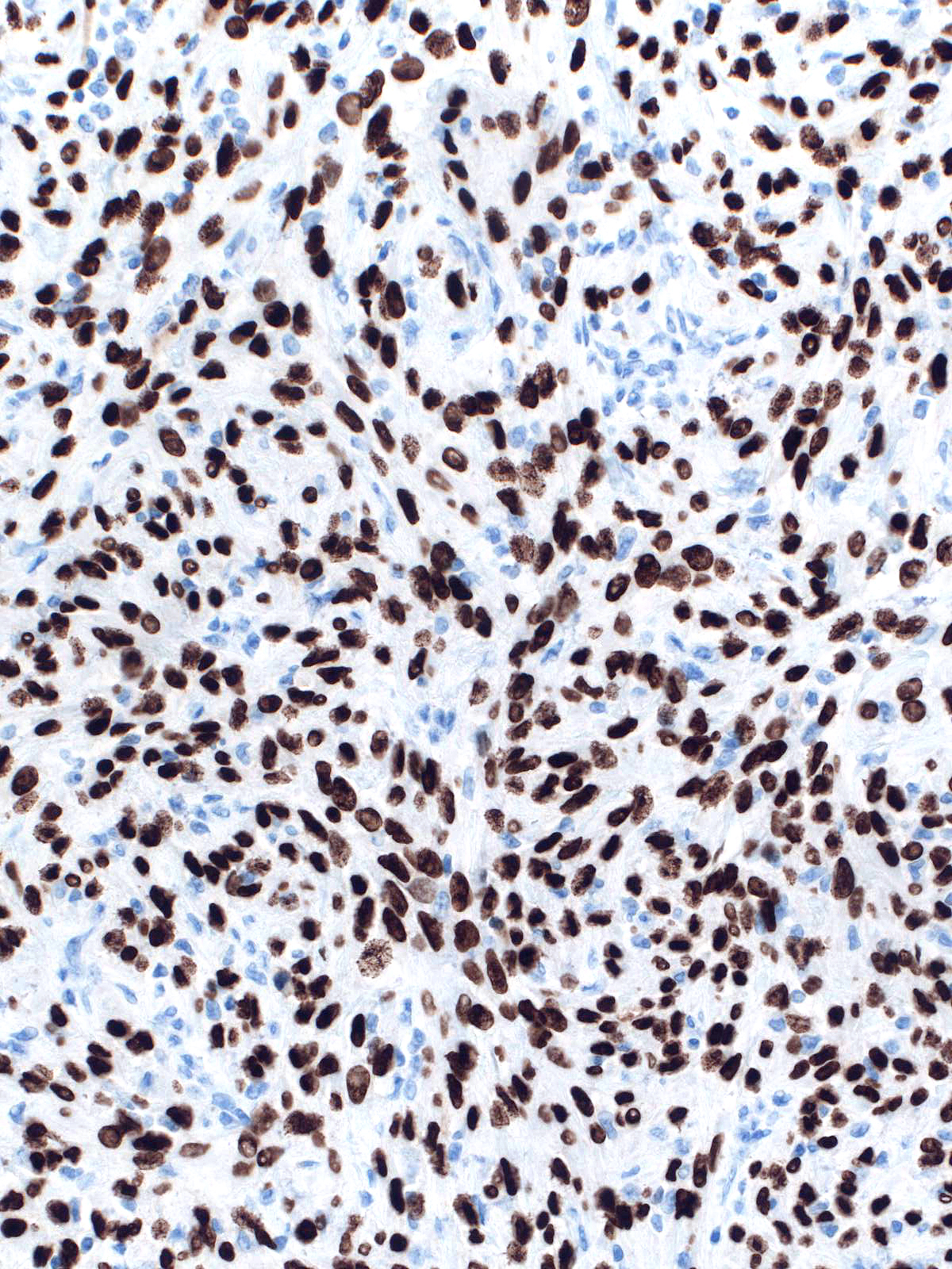
Neoplastic myoepithel cells and the cells of spindle cell carcinoma regularly stain for p63.
 |
 |
Smooth Muscle Myosin Heavy Chain
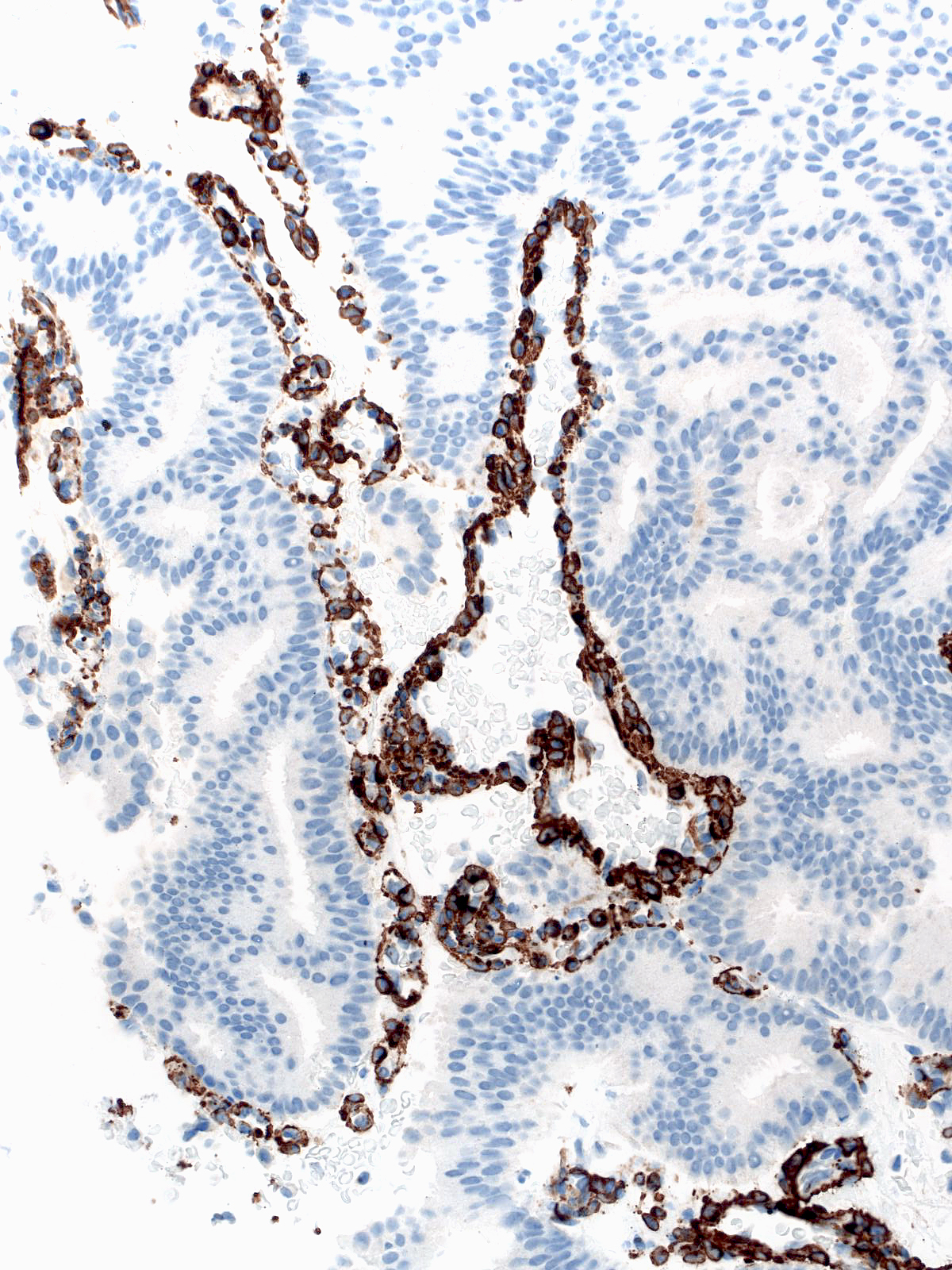
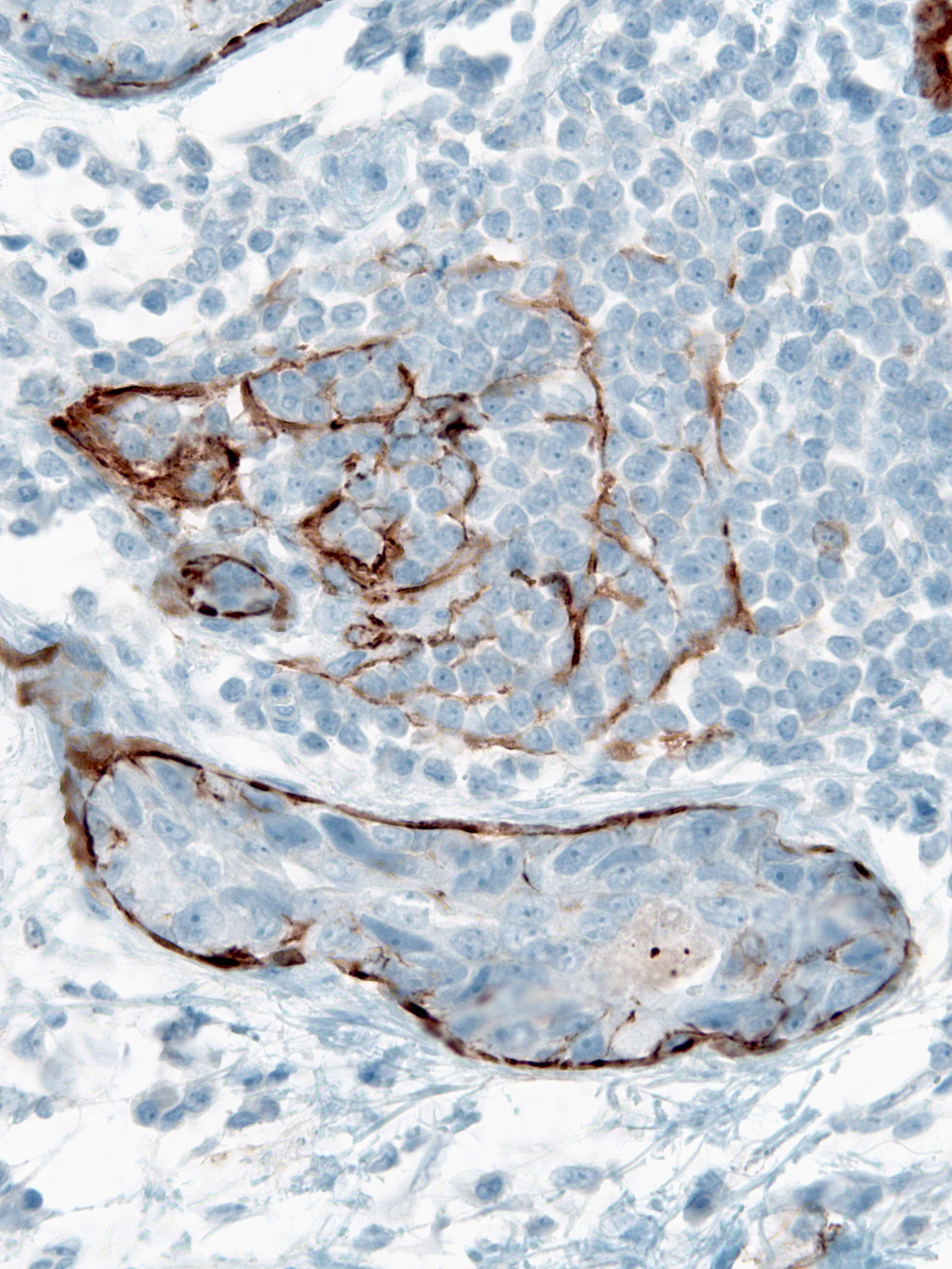
Several types of cells other than myoepithelial cells express this protein. Pay attention to the features evident on the H&E-stained sections to avoid misinterpretations.
 |
 |
Calponin
Antibodies to calponin frequently react with stroma myofibroblasts. When the myofibroblasts sit next to epithelial cells, one could mistake the stromal cells for myoepithelial cells and thereby fail to recognize the invasive nature of carcinoma cells.
| Staining of stromal cells could lead you to mistake this small cluster of invasive carcinoma cells for a focus of DCIS. | 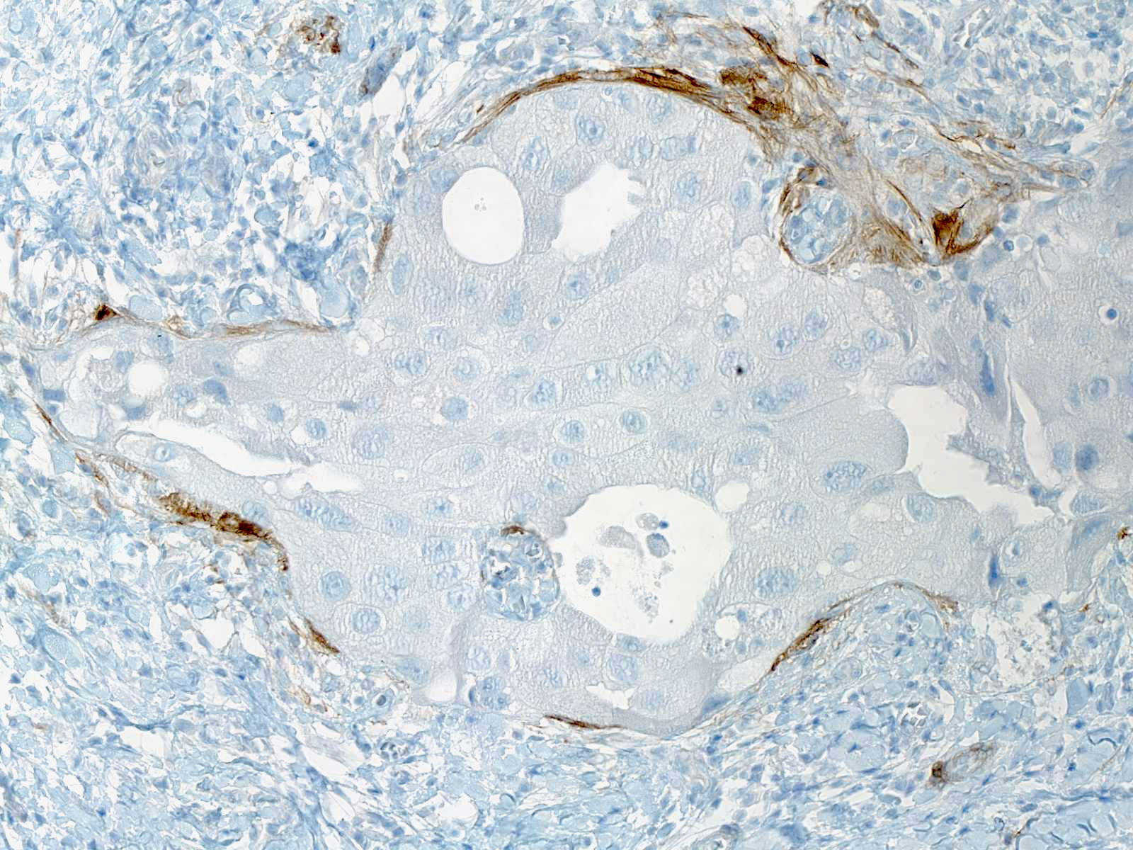 |
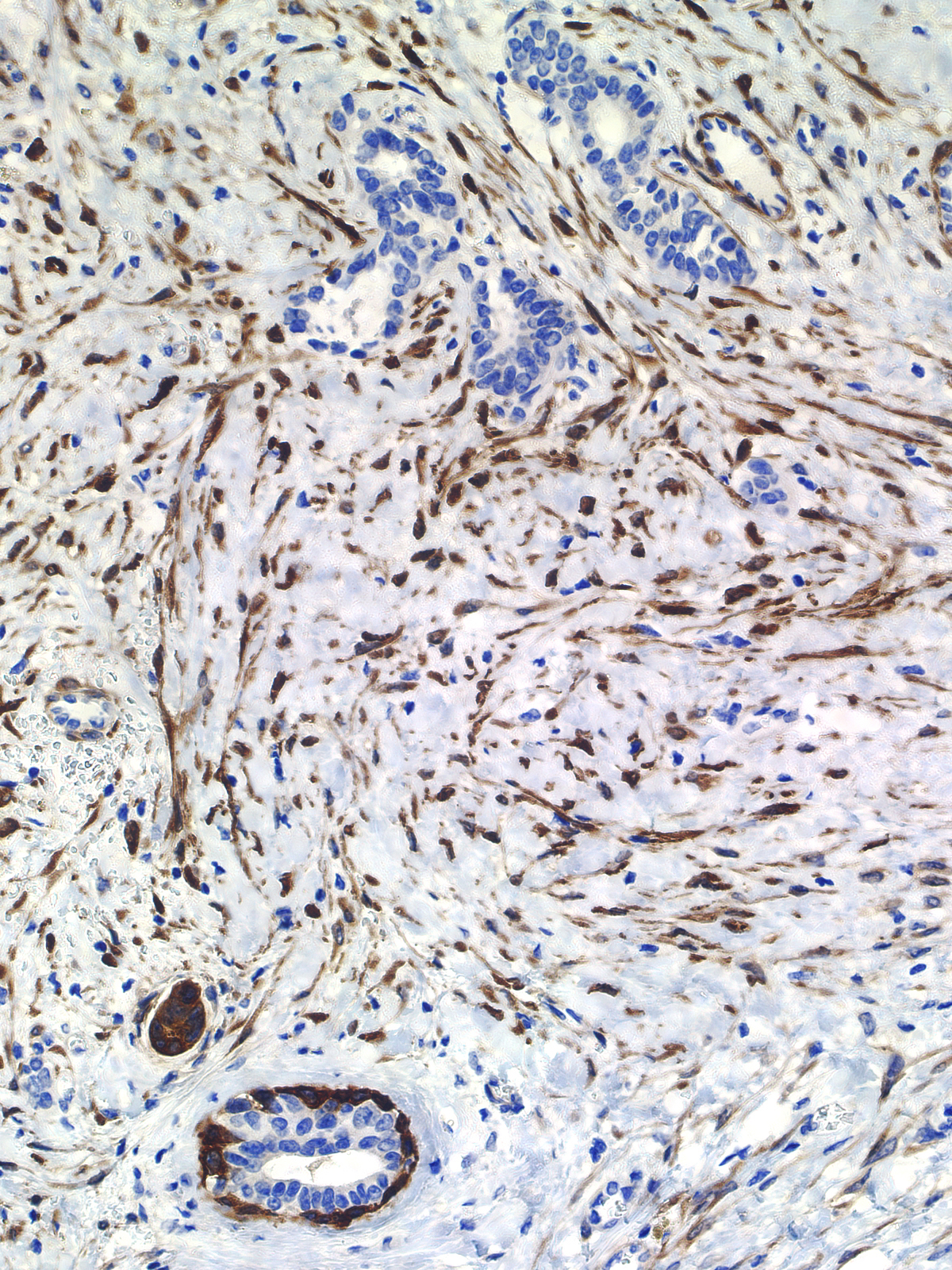
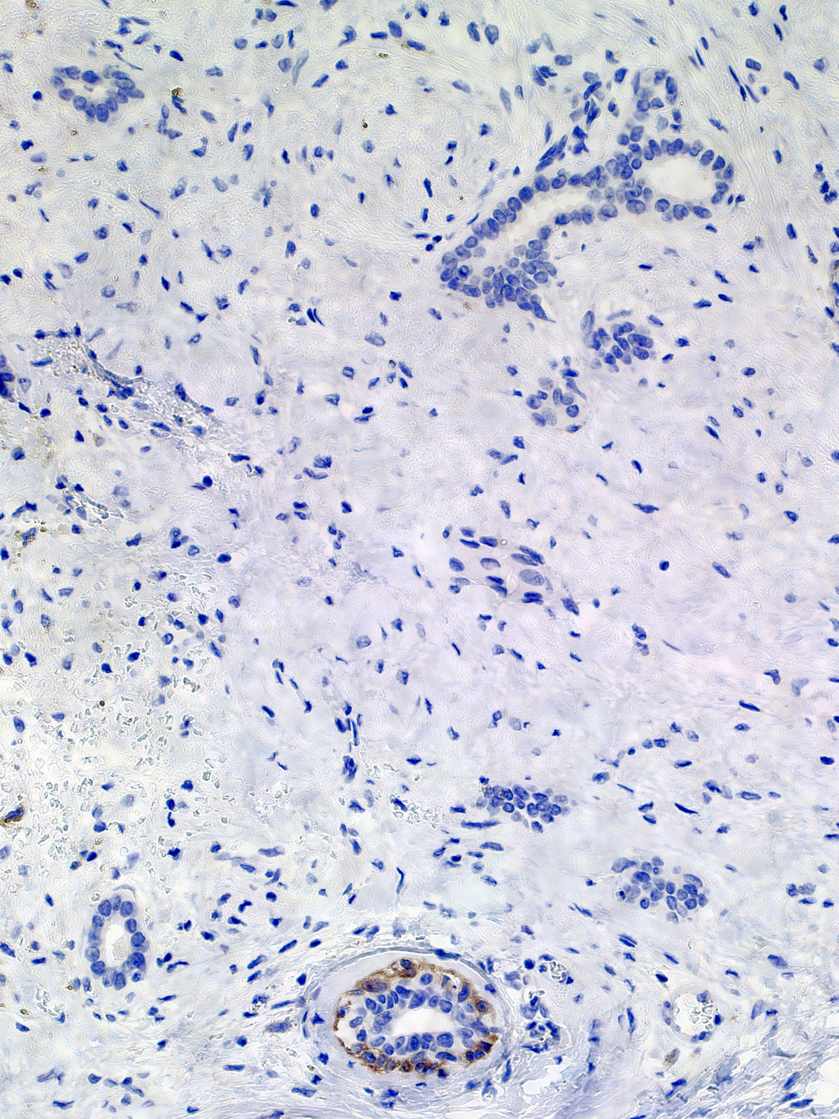
The presence of many reactive myofibroblasts can make it difficult both to detect myoepithelial cells and to appreciate the absence of myoepithelial cells. Use this stain in conjunction with other myoepithelial markers.
The staining of many stromal cells makes it difficult to appreciate the apsence of mye epithelial cells surrounding the nests in the upper portion of the field (left). The stain for smooth muscle myosin heavy chain makes it easy to recognize the absence of myoepithelial cells around the clands in question.
 |
 |
The staining of a cell for calponin does not guarantee its benign nature, for neoplastic myoepithelial cells can stain for the protein
| The peripheral cells in this adenosquamous carcinoma stain for calponin in a pattern similar to that seen in normal glands | 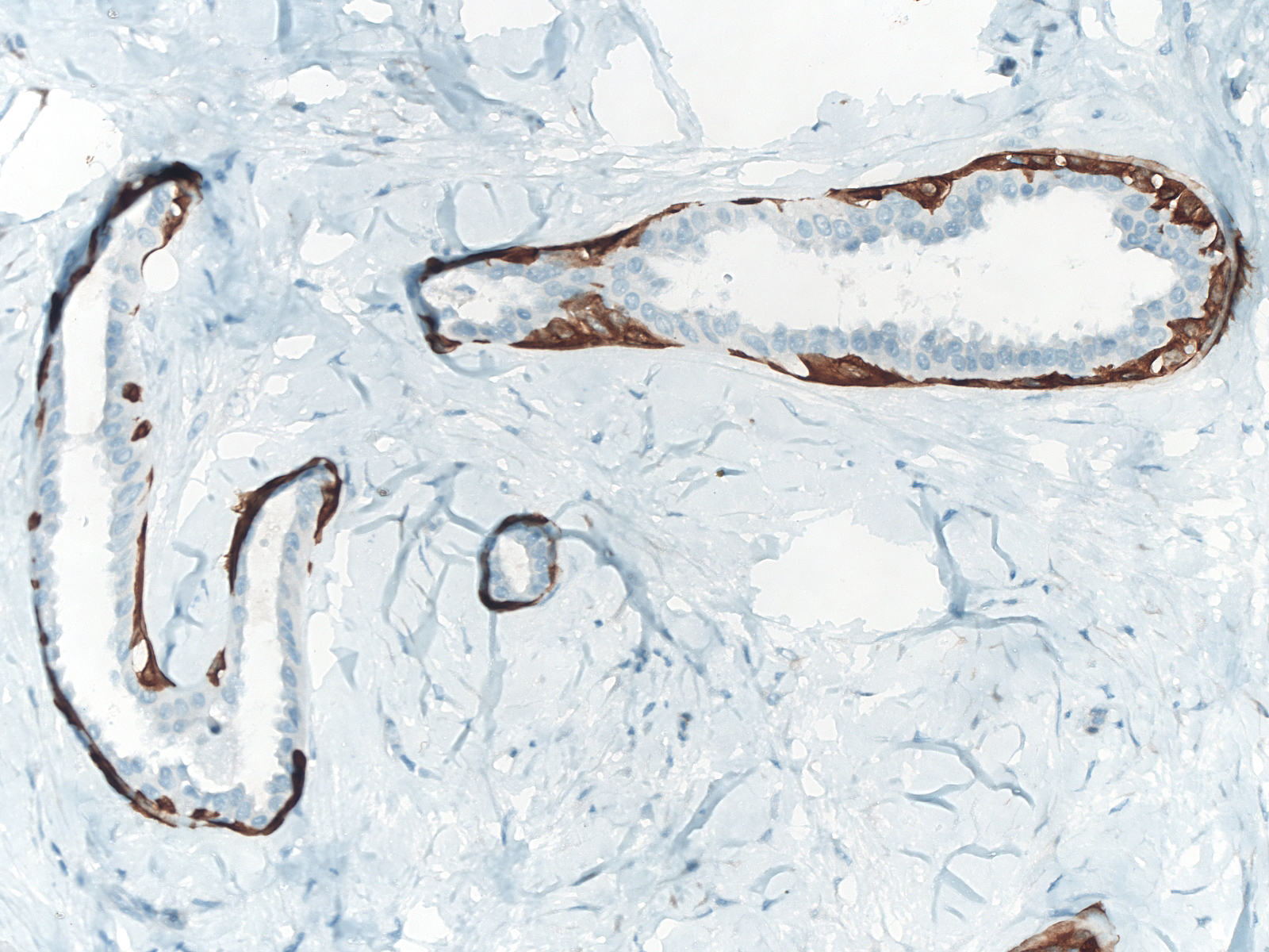 |
D2-40
Do not confuse the granular staining of myoepithelial cells with the linear staining of lymphatic endothelial cells.
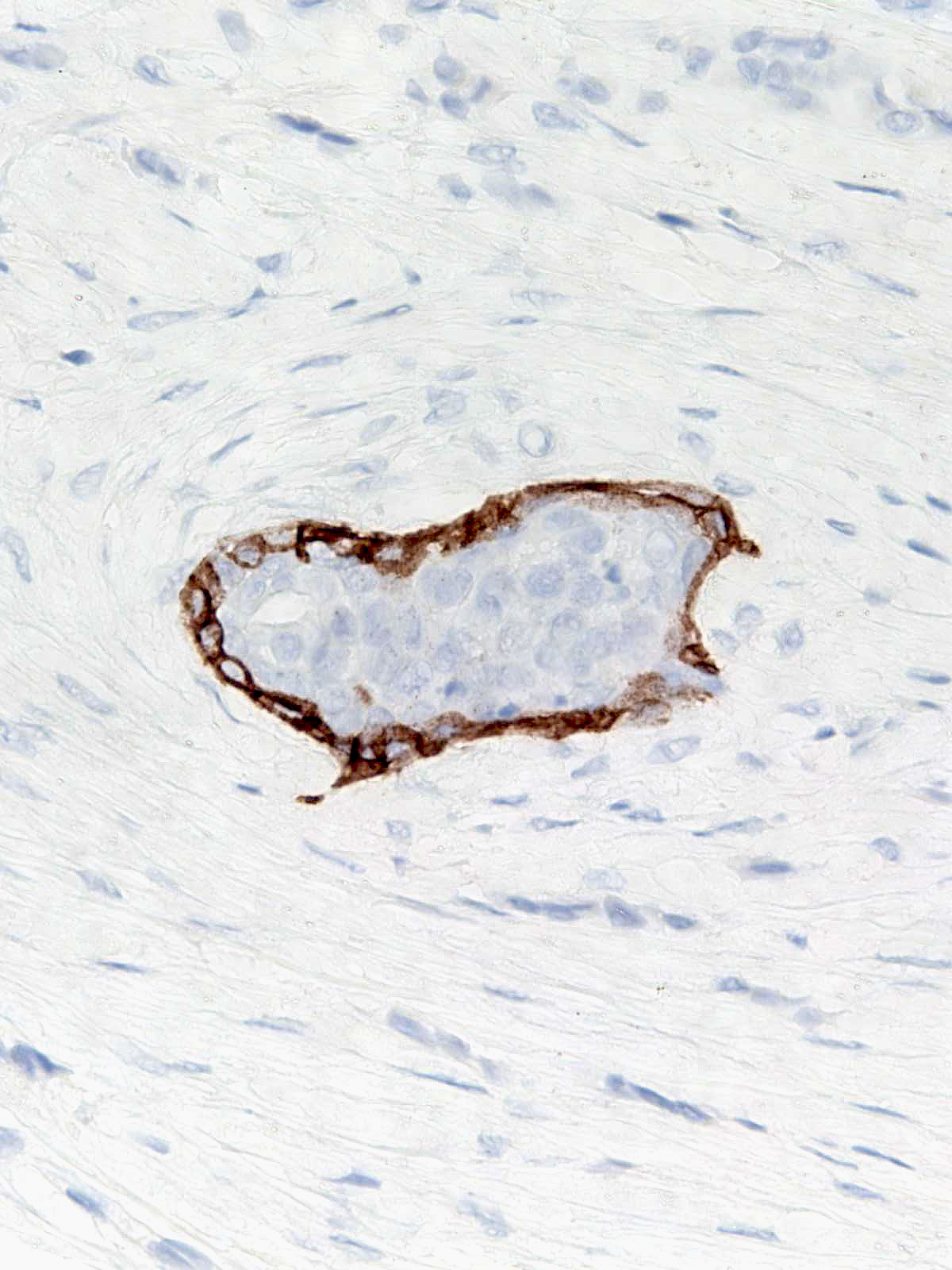 |
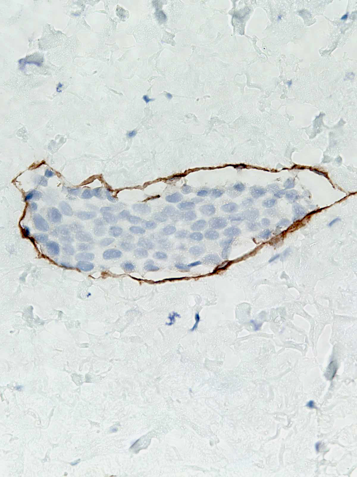 |