Difference between revisions of "Breast: Normal Anatomy of the Breast"
| (25 intermediate revisions by the same user not shown) | |||
| Line 1: | Line 1: | ||
| − | {{:TOC}} | + | {{:TOC}}{{img3|1-1 NA normal nipple 6.30.14-2.jpg|Cross section of a normal nipple and ducts|}} |
| − | |||
== Location == | == Location == | ||
| − | Breast tissue occasionally forms beyond the confines of the gland itself. This ectopic tissue can occur anywhere in the milk line, a region that runs from the axilla to the groin. The misplaced tissue can create an accessory nipple or a mass composed of primitive breast tissue. The ectopic tissue can show physiological changes and can give rise to mammary lesions such as pseudoangiomatous stromal hyperplasia, fibroadenomas, and carcinomas. | + | {{img1|Breast tissue occasionally forms beyond the confines of the gland itself. This ectopic tissue can occur anywhere in the milk line, a region that runs from the axilla to the groin. The misplaced tissue can create an accessory nipple or a mass composed of primitive breast tissue. The ectopic tissue can show physiological changes and can give rise to mammary lesions such as pseudoangiomatous stromal hyperplasia, fibroadenomas, and carcinomas.|1-2 Milk lines.jpg|}} |
| − | |||
== Glandular Tissue == | == Glandular Tissue == | ||
| − | Mammary glandular tissue consists of arborizing ducts and terminal lobules. The largest ducts, termed lactiferous sinuses, drain onto the skin of the nipple. They number between 15 and 25, have diameters of a few millimeters, and display folded, stellate configurations when cut in cross section. The lactiferous sinuses form a compact bundle in the nipple and extend posteriorly to become large ducts. As the large ducts ramify within the gland, they diminish in caliber and branch to form small ducts, ductules, and extralobular terminal ductules, which together comprise the conducting portion of the mammary glandular tree. The extralobular terminal ductules drain terminal duct lobular units. The secretory component of the gland consists of intralobular terminal ductules and their attached acini. | + | {{img1|Mammary glandular tissue consists of arborizing ducts and terminal lobules. The largest ducts, termed lactiferous sinuses, drain onto the skin of the nipple. They number between 15 and 25, have diameters of a few millimeters, and display folded, stellate configurations when cut in cross section. The lactiferous sinuses form a compact bundle in the nipple and extend posteriorly to become large ducts. As the large ducts ramify within the gland, they diminish in caliber and branch to form small ducts, ductules, and extralobular terminal ductules, which together comprise the conducting portion of the mammary glandular tree. The extralobular terminal ductules drain terminal duct lobular units. The secretory component of the gland consists of intralobular terminal ductules and their attached acini.|1-3 NA normal-lobule-cooper 7.29.14.jpg|}} |
| − | + | {{img4|1-4 NA_lactiferous_sinuses_6.30.14.jpeg|1-5 NA_large_ducts_6.30.14.jpeg|Lactiferous sinuses connect the breast glandular tissue with the skin of the nipple.|Large ducts branch and narrow, forming the main conductive pathways.}} | |
| + | {{img4|1-6 NA_duct_lobule_6.30.14.jpeg|1-7 NA_Ductule_TDLU_6.30.14.jpeg.jpg|Small duct and lobules|Ductule and terminal duct-lobular unit (TDLU)}} | ||
| + | The epithelium that lines the glands appears similar throughout the breast. The epithelium consists of two layers of cells: luminal cells, and myoepithelial cells. The luminal cells display polarization. They exhibit columnar or cuboid shapes and they possess oval or round nuclei, small nucleoli, and varying amounts of eosinophilic cytoplasm. The apical surface abuts the gland lumen, and the basal end attaches to the basement membrane. Luminal cells carry out secretory and absorptive functions. The myoepithelial cells appear flattened along the basement membrane. Being covered by the luminal cells, the myoepithelial cells do not touch the gland lumen. They contain small, triangular nuclei, and it is often difficult to see their cytoplasm. Myoepithelial cells form dendrite‑like cell processes, which extend along the basement membrane and sometimes among the luminal cells. They have contractile properties and they contribute to the formation and maintenance of the basement membrane. Pluripotent cells capable of differentiating into both luminal and myoepithelial cells occupy basal positions throughout the epithelium. T-cells and histiocytes often percolate among the epithelial cells. A layer of homogeneous, eosinophilic basement membrane underlies the epithelium. | ||
| + | {{img1|Normal ducts are lined by two layers of epithelial cells: outer luminal cells and inner myoepithelial cells. Basement membrane underlies the epithelium.|1-8 NA_duct_60x_6.30.14.jpeg|}} | ||
| + | {{img1|Acini are also composed of outer luminal epithelial cells and inner myoepithelial cells. These luminal epithelial cells are plump compared to the myoepithelial cells. Basement membrane enshrouds the epithelium, a thin barrier separating the epithelial cells from the numerous capillaries in the specialized stroma.|1-9 NA_acini_6.30.14.jpeg|}} | ||
| + | {{img1|During pregnancy, the terminal duct lobular units enlarge as milk-producing glands sprout from the existing acini.|1-10 NA_lactation_low_6.30.14.jpeg|}} | ||
| + | {{img1|The lumens of the milk- producing glands dilate and the luminal epithelial cells develop abundant vacuolated cytoplasm.|1-11 NA_lactation_medium_6.30.14.jpeg|}} | ||
| + | {{img1|The luminal cells of lactation feature one or two large nuclei and prominent nucleoli. Loosening of the intercellular attachments allows the cytoplasm of the luminal cells to bulge into the lumen, which also contains eosinophilic secretion and bits of sloughed cytoplasm.|1-12 NA_lactation_high_6.30.14.jpeg|}} | ||
| + | {{img1|Mammary tissue in both boys and girls appears identical until puberty. The glandular tissue of the male breast does not develop further and does not display the complexity seen in the adult female breast. Mammary glandular tissue in men consists only of simply branching ducts, although in uncommon situations very simple lobules can form.|1-13 NA_male_6.30.14.jpeg|}} | ||
| + | == Stroma == | ||
| + | Stroma is found internal to the basement membrane and epithelium. There are three types of mammary stroma: nipple stroma; specialized/intralobular stroma; and nonspecialized/extralobular stroma. | ||
| + | {{img4|1-14 NA_Muscle_bundles_6.30.14.jpeg.jpg|1-15 Nipple_MS17-2097_5_x_B.jpg|1-16 Nipple_D2-40_MS17-2097_D2-40_5_x_A.jpg| | ||
| + | Fascicles of smooth muscle running haphazard among collagen bundles, capacious thin walled blood vessels, and abundant lymphatic vessels characterize the stroma of the nipple.|Nipple stroma: the capacious thin walled blood vessels and lymphatics are especially apparent by D2-40 immunostain.||}} | ||
| + | Specialized stroma (also called intralobular stroma) forms a layer around the entire mammary glandular tree. This type of stroma appears most obvious within lobules, where it consists of slender bundles of collagen, fibroblasts, and capillaries embedded in blue grey, myxoid ground substances. One can also observe plasma cells, lymphocytes and histiocytes in the specialized stroma. In most cases, one can barely discern the specialized stroma around ducts; however, on close inspection a concentric arrangement of loose collagen bundles and a ring of capillaries combined with scattered chronic inflammatory cells mark the periductal specialized stroma. | ||
| + | {{img1|Easily recognized in-between the acini in this normal lobule, specialized stroma consists of slender bundles of collagen, fibroblasts, and capillaries in a blue-grey extracellular matrix.|1-17 NA_normal_lobule_6.30.14.jpeg|}} | ||
| + | {{img1|A modest amount of specialized stroma surrounds normal ducts. Click the picture to enlarge and notice the ring of capillaries and concentric bundles of collagen marking the specialized stroma.|1-18 NA_periductal_stroma_6.30.14.jpeg|}} | ||
| + | Nonspecialized stroma (also called extralobular stroma) surrounds the specialized stroma of ducts and ductules and encloses entire lobules. The nonspecialized stroma provides the structure of the breast and supports the function of the glandular tissue. Nonspecialized stroma consists of bundles of dense, eosinophilic collagen with intermixed large and small blood vessels, lymphatic vessels, fibroblasts, and myofibroblasts. Anteriorly, the nonspecialized stroma blends with the fibrous tissue of the reticular dermis and posteriorly it blends with the prepectoral fascia. Fibrous bands extending anteriorly from the prepectoral fascia are referred to as Cooper's ligaments. The fibrous connective tissue of the nonspecialized stroma intermixes with adipose tissue. | ||
| + | {{img1|An anterior view of the fibrous tissue which supports the breast and envelopes the glandular network. The nipple is seen in the top center of the image. (Cooper, 1840)|1-19 NA_cooper-ligaments_7.29.14.jpg|}} | ||
| + | {{img1|Section through the nipple showing the glandular network and suspensory fibrous tissue. The fibrous tissue forms two partitions, anchoring the gland anteriorly to the skin and posteriorly to the pectoral muscle. (Cooper, 1840)|1-20 NA_fibrous-tissue_7.29.14.jpg|}} | ||
| + | {{img1|During development, the primitive epidermis of the anterior chest invaginates into the mesoderm to create the basic structure of the breast's glandular component. Despite the complexity of the final structure, the breast maintains its layered compartments, which could be stretched out to reveal flat sheets that never intermix. The layers of the breast from most external to most internal are: (1) luminal epithelial cells; (2) myoepithelial cells; (3) basement membrane; (4) specialized stroma; (5) non-specialized stroma; and (6) fat. Mixing of non-juxtaposed layers likely represents a pathologic process.|1-21 layers-2.jpg|}} | ||
| + | == Lymphoid Tissue == | ||
| + | The breast contains mucosa associated lymphoid tissue. T-cell aggregates occupy the specialized stroma around the walls of large ducts and occasionally infiltrates the overlying epithelium, which consequently appears hyperplastic. These T-cells probably serve to detect offending antigens. The specialized stroma of lobules also contains plasma cells and their precursors. The plasma cells produce the secretory IgA present in milk. Collections of B-cells may encircle small veins. | ||
| + | {{img4|1-22 Lymphoid_aggregate_5_x.jpg|1-23 Lymphoid_aggregate_10_x.jpg|Mucosa associated lymphoid tissue: T-cells form aggregates in the specialized stroma around large ducts.|The epithelium overlying such lymphoid aggregates can appear hyperplastic.}} | ||
| + | {{img4|1-24 Lobule_w_plasma_cells.jpg|1-25 Perivasc_lymphs.jpg|Plasma cells in the specialized stroma of a lobule|Perivascular B lymphocytes}} | ||
| + | == Skin == | ||
| + | The skin covering the breast resembles the skin covering the remainder of the body. It consists of stratified squamous epithelium and the usual adnexal structures. Like other apocrine-gland-bearing skin, mammary epithelium contains Toker cells. These cells are found near the orifices of the lactiferous sinuses and other ducts that drain onto the skin of the nipple and areola. They sit in the basal region of the epithelium and occur singly and in small groups. Toker cells appear about the same size as their neighboring keratinocytes and they contain bland nuclei, small nucleoli, and pale, finely eosinophilic cytoplasm. Toker cells stain for CK7, but they do not stain for Her2. Toker cells may give rise to the malignant cells in certain cases of Paget disease of the breast. The lack of nuclear pleomorphism, the restriction to the basal aspect of the epidermis, and the absence of staining for HER2 help to distinguish Toker cells from the carcinoma cells of Paget disease. | ||
| + | {{img1|Toker cells are basally oriented, about the same size as keratinocytes, contain bland nuclei, and stain for keratin 7 but not Her2.|1-26 Toker_40_x_E.jpg||}} | ||
== Quiz == | == Quiz == | ||
<bootstrap_collapse text="Start Quiz"><p><https://hub.partners.org/pathology/assessment/instructions?assessment_id=16607586" height="666" width="100%"></htmltag> | <bootstrap_collapse text="Start Quiz"><p><https://hub.partners.org/pathology/assessment/instructions?assessment_id=16607586" height="666" width="100%"></htmltag> | ||
Latest revision as of 12:37, July 8, 2020
 |
||
Location
| Breast tissue occasionally forms beyond the confines of the gland itself. This ectopic tissue can occur anywhere in the milk line, a region that runs from the axilla to the groin. The misplaced tissue can create an accessory nipple or a mass composed of primitive breast tissue. The ectopic tissue can show physiological changes and can give rise to mammary lesions such as pseudoangiomatous stromal hyperplasia, fibroadenomas, and carcinomas. | 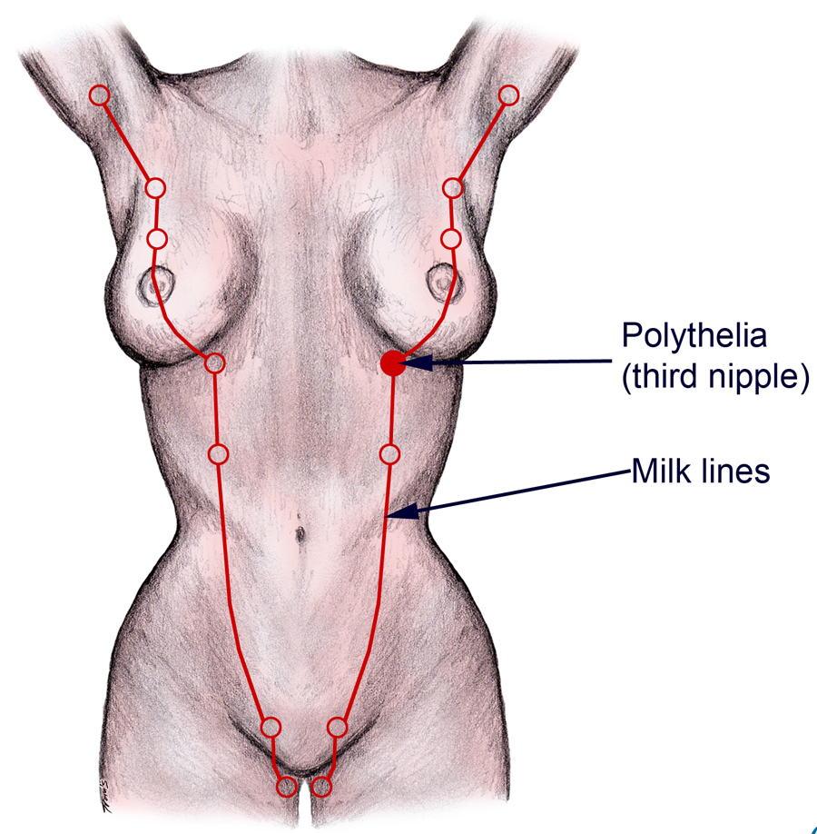 |
Glandular Tissue
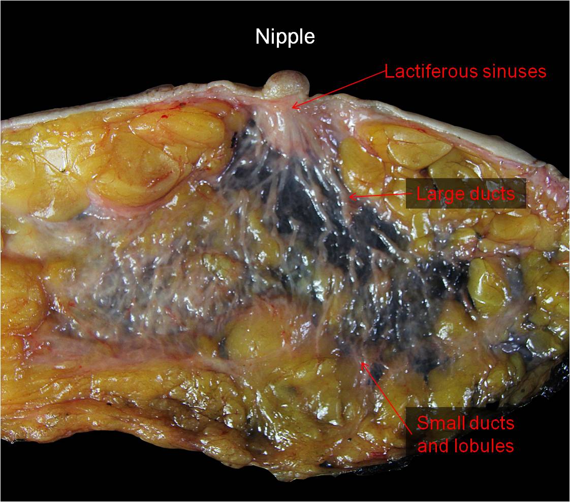
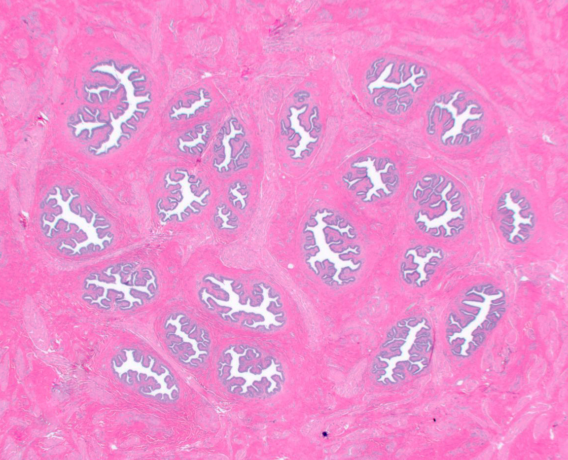
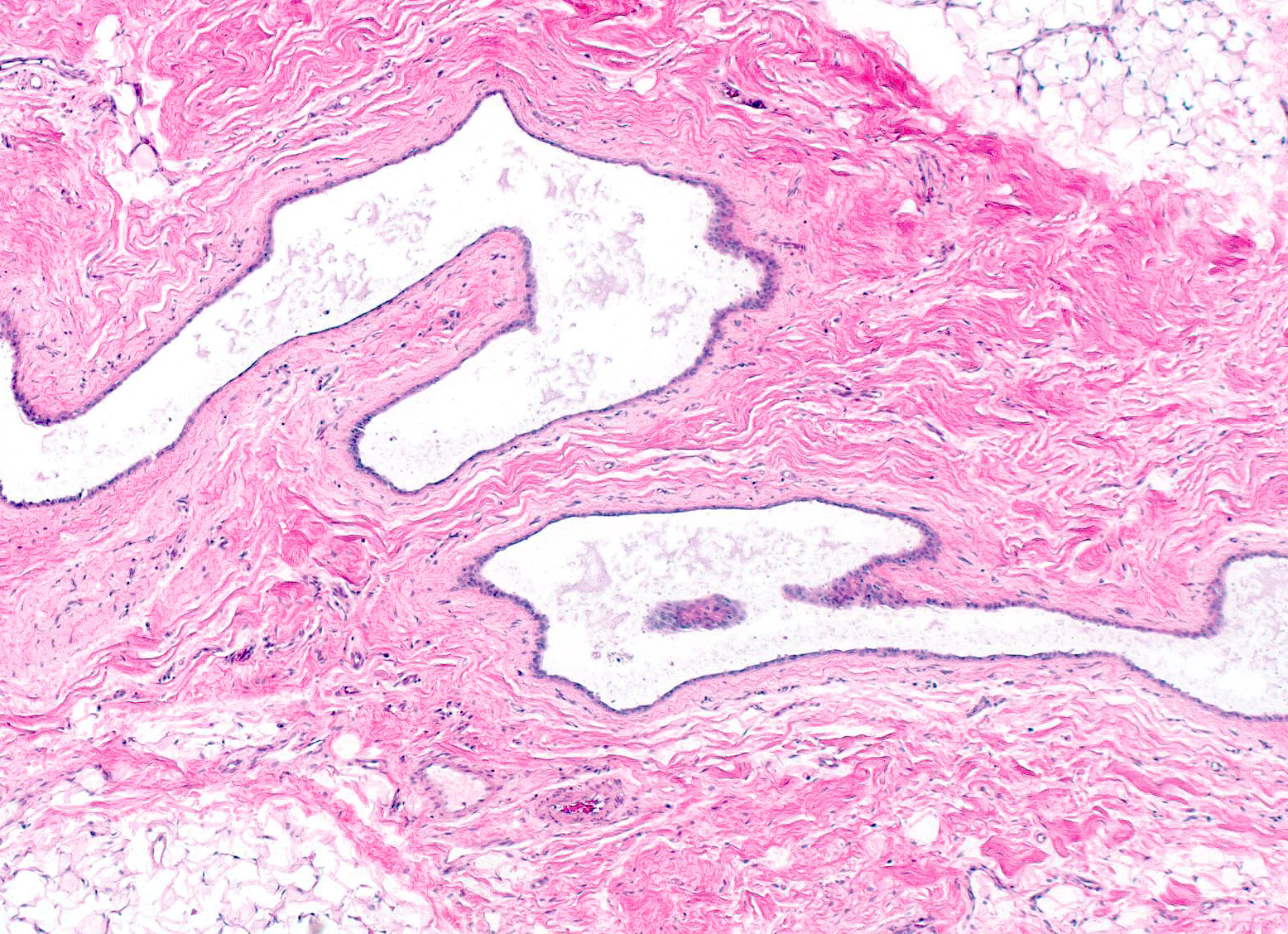
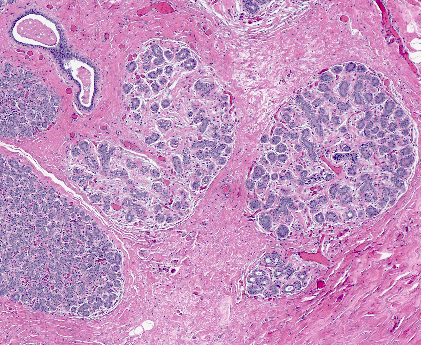
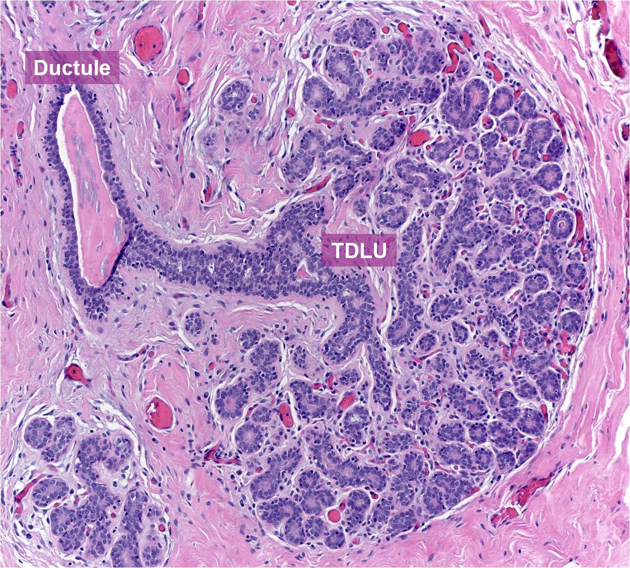
| Mammary glandular tissue consists of arborizing ducts and terminal lobules. The largest ducts, termed lactiferous sinuses, drain onto the skin of the nipple. They number between 15 and 25, have diameters of a few millimeters, and display folded, stellate configurations when cut in cross section. The lactiferous sinuses form a compact bundle in the nipple and extend posteriorly to become large ducts. As the large ducts ramify within the gland, they diminish in caliber and branch to form small ducts, ductules, and extralobular terminal ductules, which together comprise the conducting portion of the mammary glandular tree. The extralobular terminal ductules drain terminal duct lobular units. The secretory component of the gland consists of intralobular terminal ductules and their attached acini. | 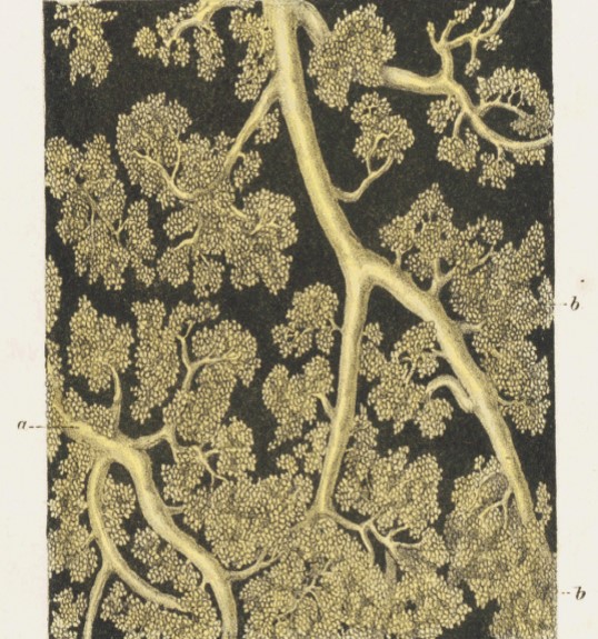 |
 |
 |
 |
 |
The epithelium that lines the glands appears similar throughout the breast. The epithelium consists of two layers of cells: luminal cells, and myoepithelial cells. The luminal cells display polarization. They exhibit columnar or cuboid shapes and they possess oval or round nuclei, small nucleoli, and varying amounts of eosinophilic cytoplasm. The apical surface abuts the gland lumen, and the basal end attaches to the basement membrane. Luminal cells carry out secretory and absorptive functions. The myoepithelial cells appear flattened along the basement membrane. Being covered by the luminal cells, the myoepithelial cells do not touch the gland lumen. They contain small, triangular nuclei, and it is often difficult to see their cytoplasm. Myoepithelial cells form dendrite‑like cell processes, which extend along the basement membrane and sometimes among the luminal cells. They have contractile properties and they contribute to the formation and maintenance of the basement membrane. Pluripotent cells capable of differentiating into both luminal and myoepithelial cells occupy basal positions throughout the epithelium. T-cells and histiocytes often percolate among the epithelial cells. A layer of homogeneous, eosinophilic basement membrane underlies the epithelium.
| Normal ducts are lined by two layers of epithelial cells: outer luminal cells and inner myoepithelial cells. Basement membrane underlies the epithelium. | 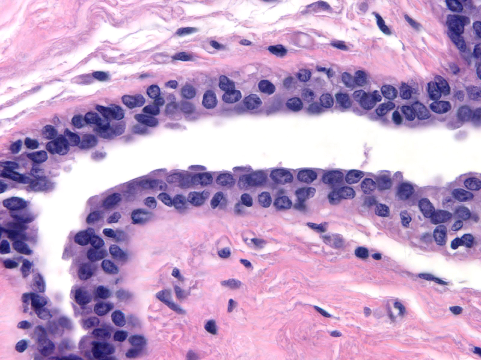 |
| Acini are also composed of outer luminal epithelial cells and inner myoepithelial cells. These luminal epithelial cells are plump compared to the myoepithelial cells. Basement membrane enshrouds the epithelium, a thin barrier separating the epithelial cells from the numerous capillaries in the specialized stroma. | 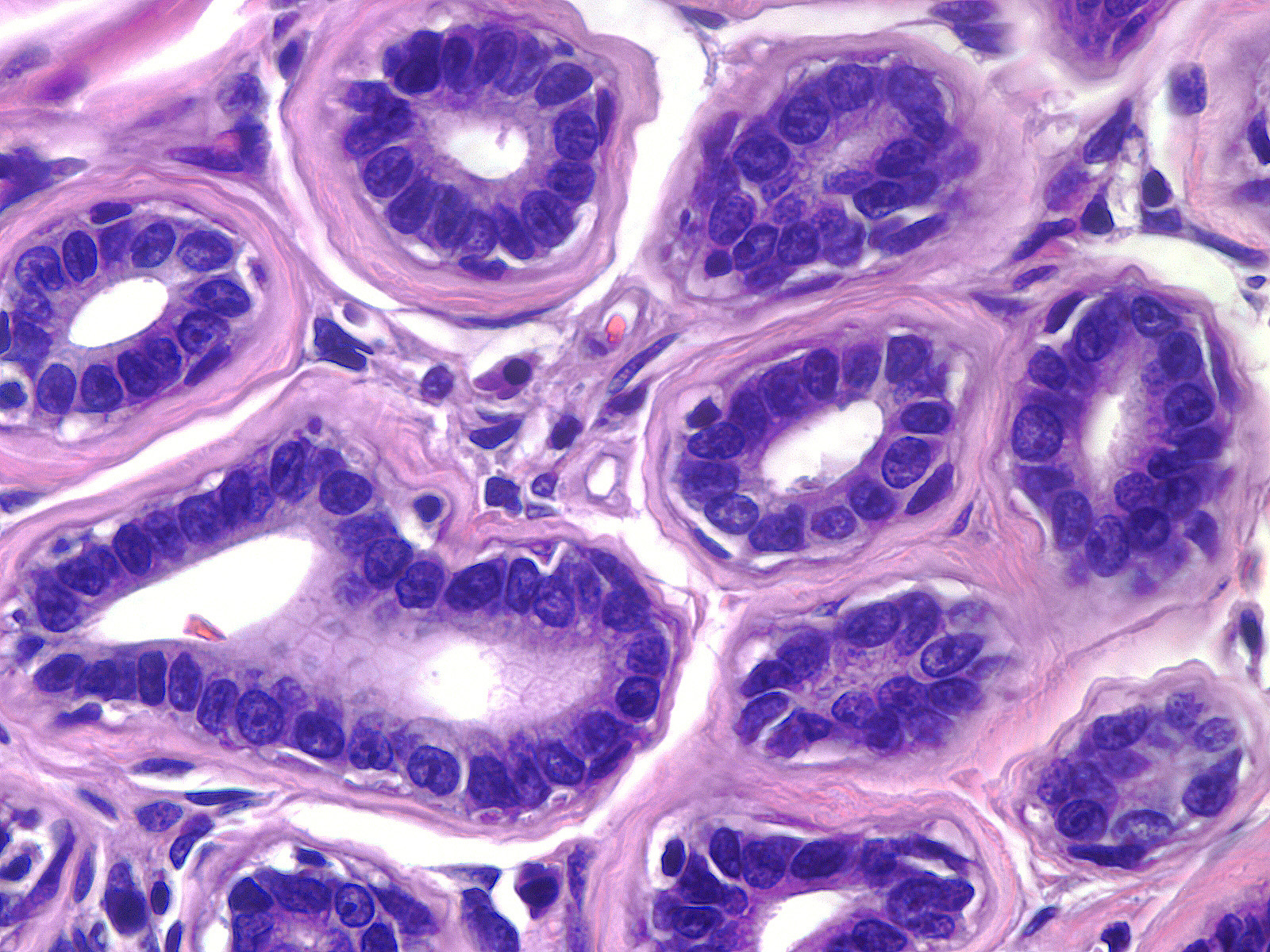 |
| During pregnancy, the terminal duct lobular units enlarge as milk-producing glands sprout from the existing acini. | 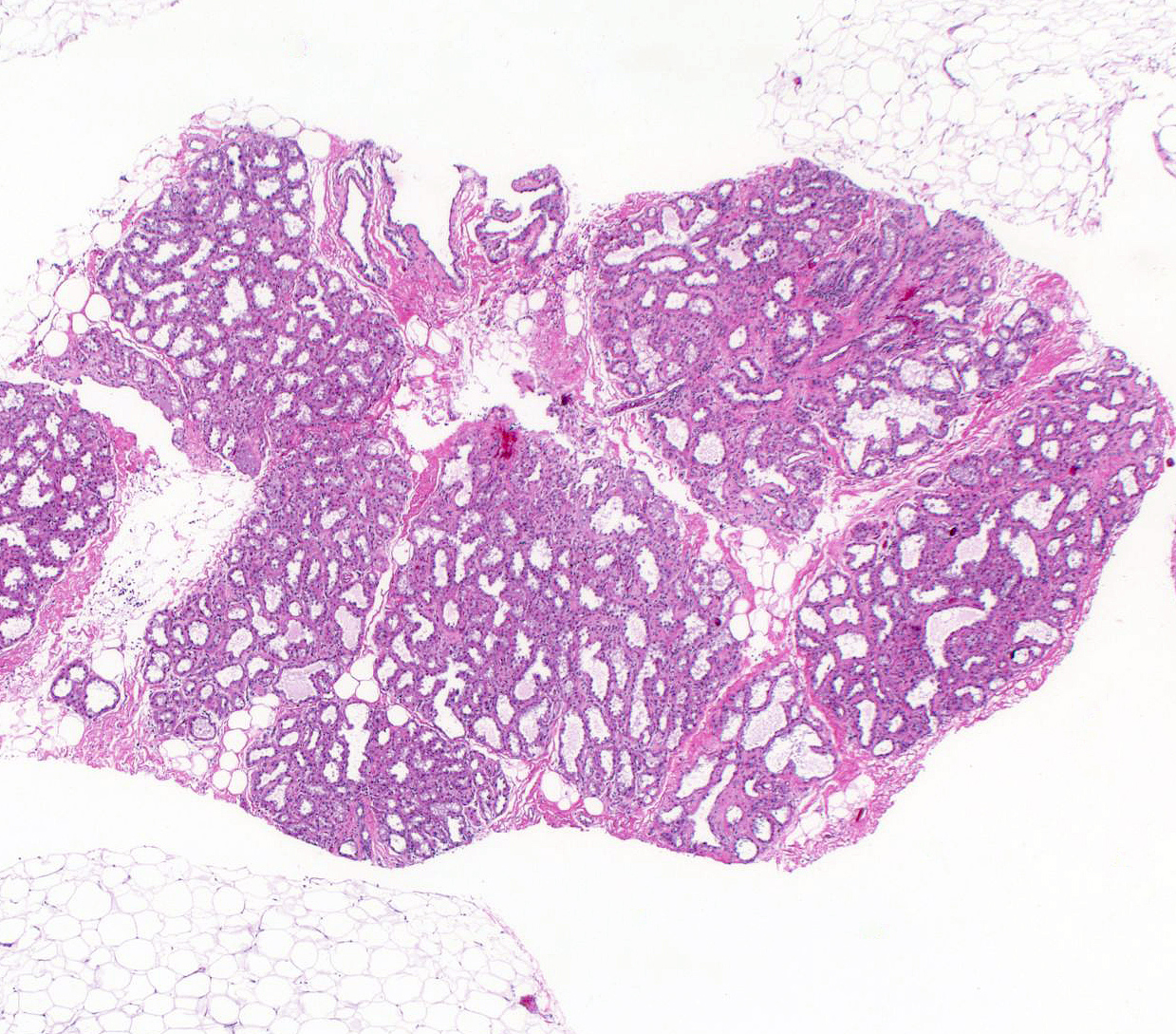 |
| The lumens of the milk- producing glands dilate and the luminal epithelial cells develop abundant vacuolated cytoplasm. | 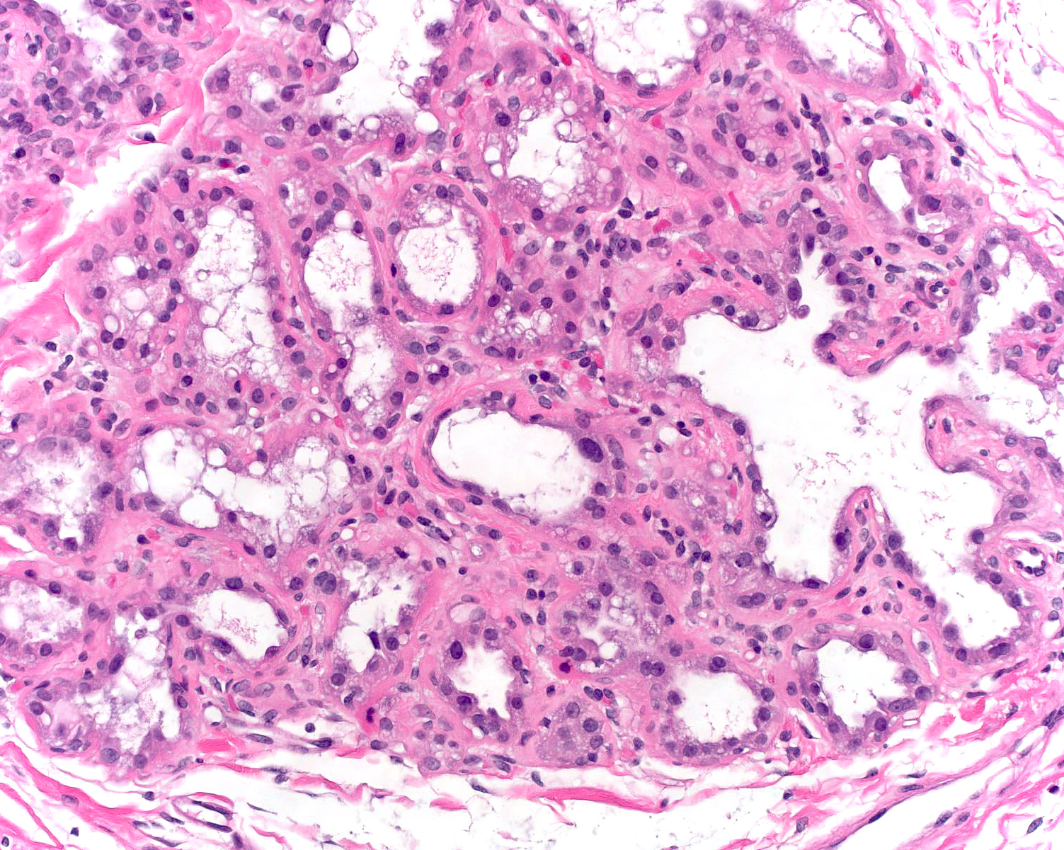 |
| The luminal cells of lactation feature one or two large nuclei and prominent nucleoli. Loosening of the intercellular attachments allows the cytoplasm of the luminal cells to bulge into the lumen, which also contains eosinophilic secretion and bits of sloughed cytoplasm. | 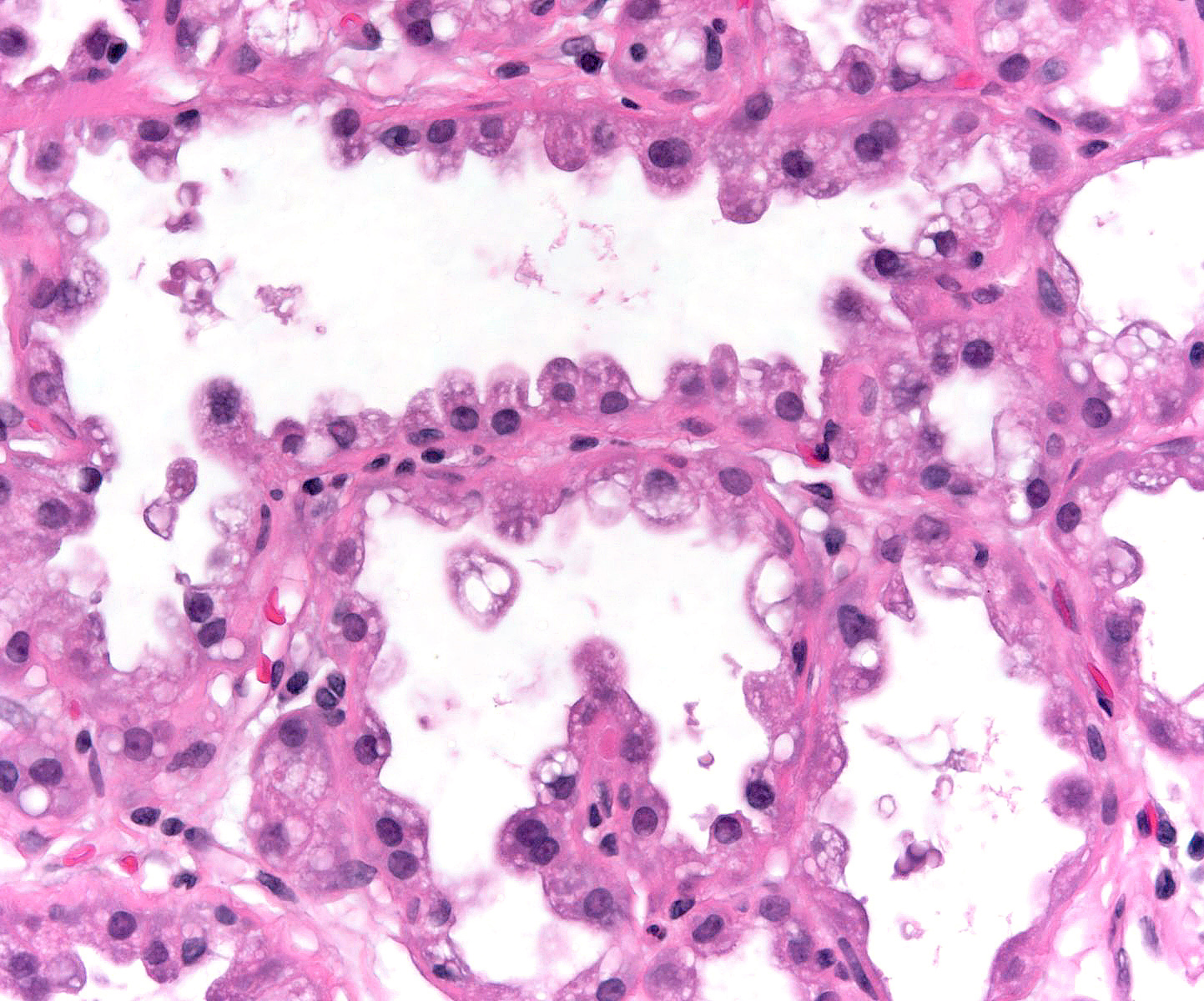 |
| Mammary tissue in both boys and girls appears identical until puberty. The glandular tissue of the male breast does not develop further and does not display the complexity seen in the adult female breast. Mammary glandular tissue in men consists only of simply branching ducts, although in uncommon situations very simple lobules can form. | 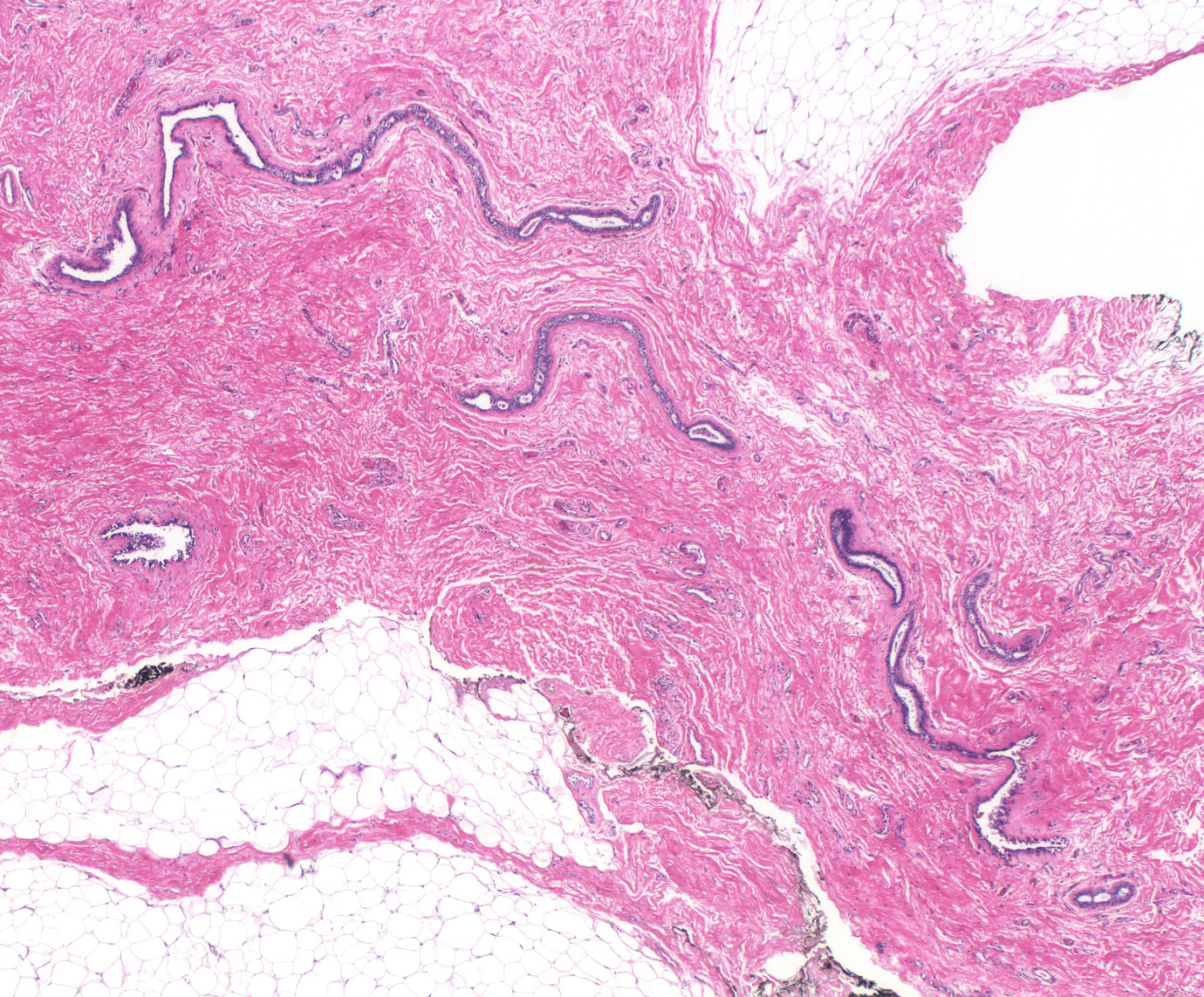 |
Stroma
Stroma is found internal to the basement membrane and epithelium. There are three types of mammary stroma: nipple stroma; specialized/intralobular stroma; and nonspecialized/extralobular stroma.
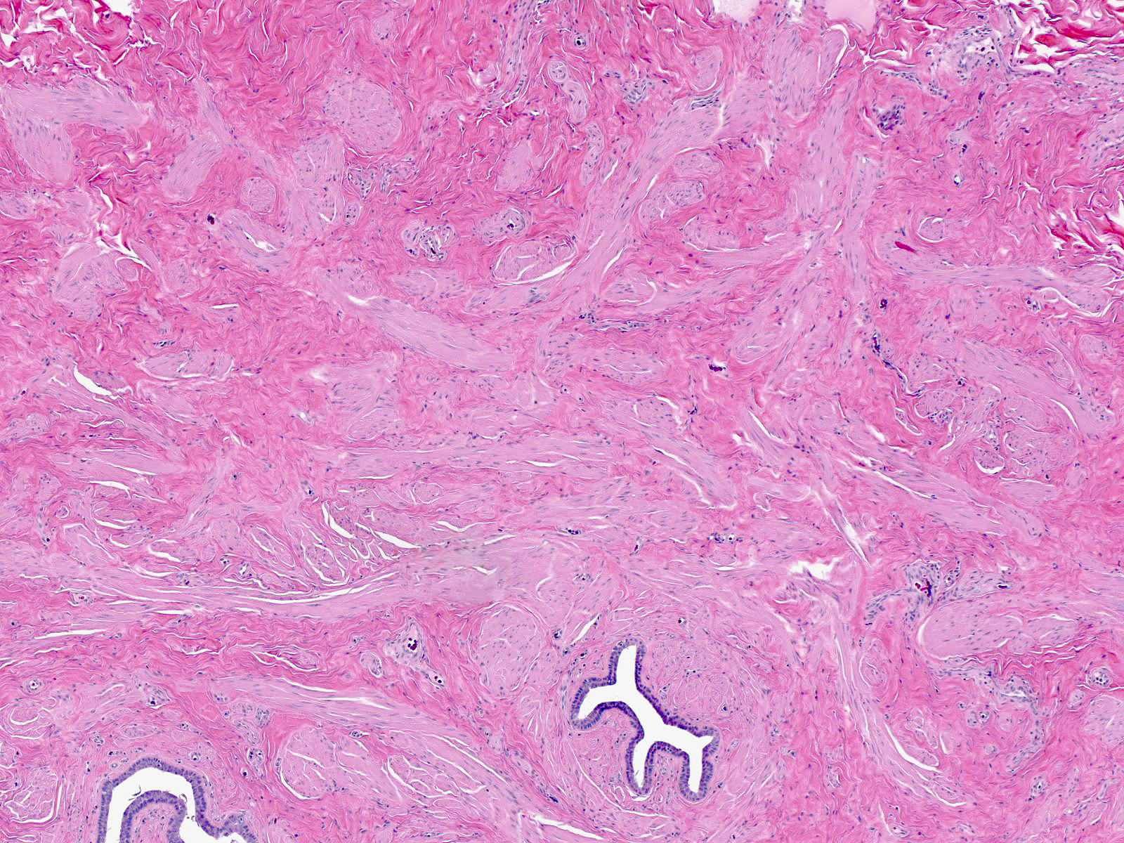 |
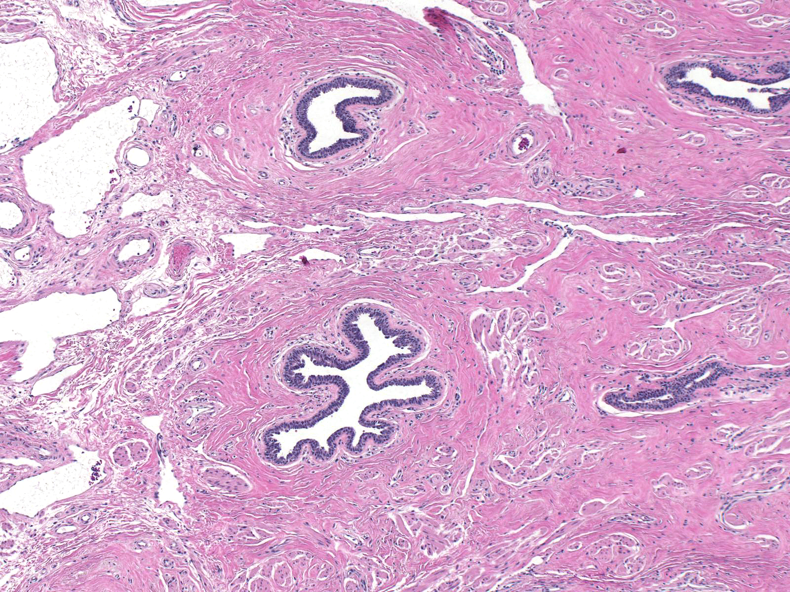 |
Specialized stroma (also called intralobular stroma) forms a layer around the entire mammary glandular tree. This type of stroma appears most obvious within lobules, where it consists of slender bundles of collagen, fibroblasts, and capillaries embedded in blue grey, myxoid ground substances. One can also observe plasma cells, lymphocytes and histiocytes in the specialized stroma. In most cases, one can barely discern the specialized stroma around ducts; however, on close inspection a concentric arrangement of loose collagen bundles and a ring of capillaries combined with scattered chronic inflammatory cells mark the periductal specialized stroma.
| Easily recognized in-between the acini in this normal lobule, specialized stroma consists of slender bundles of collagen, fibroblasts, and capillaries in a blue-grey extracellular matrix. | 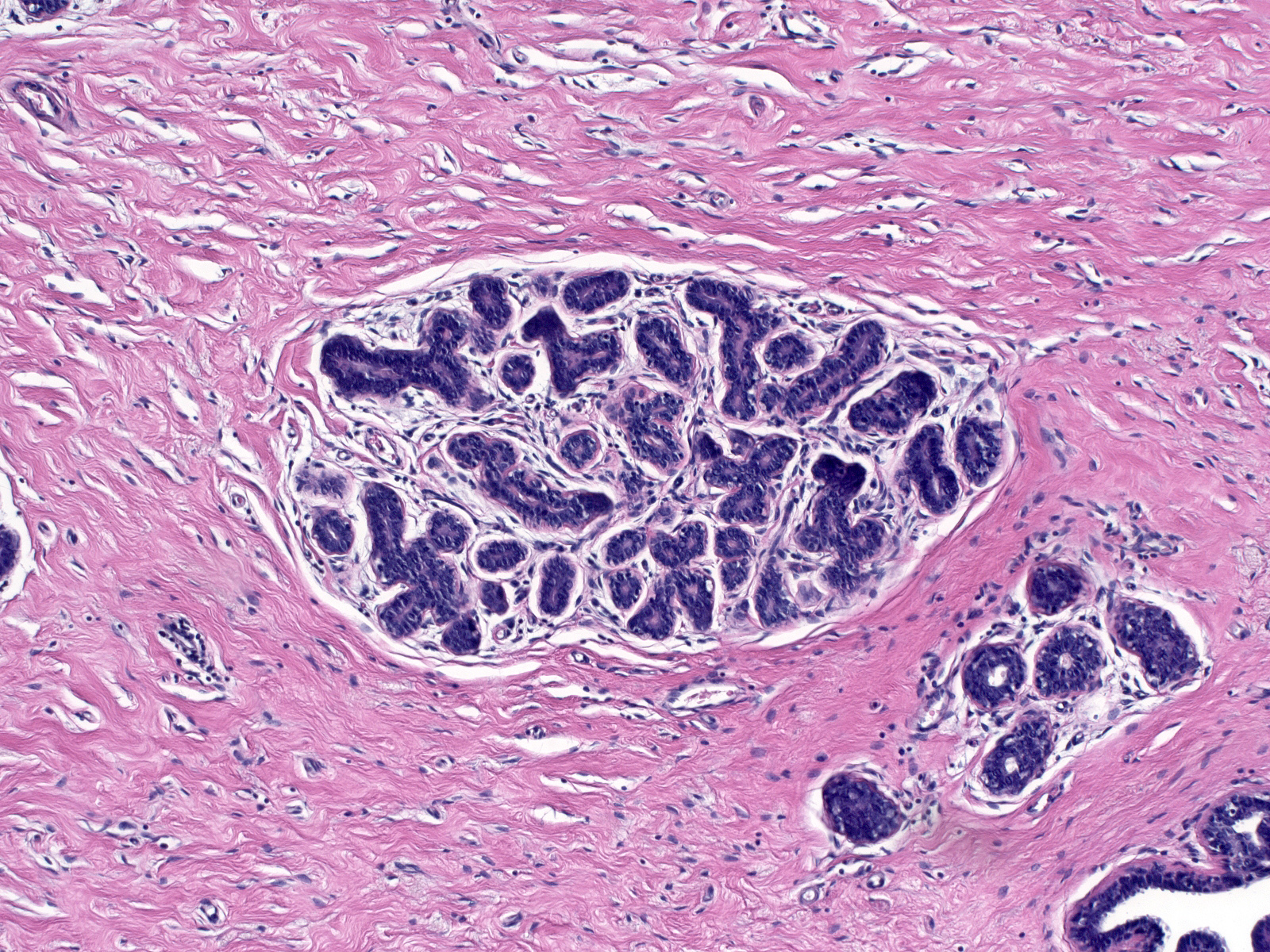 |
| A modest amount of specialized stroma surrounds normal ducts. Click the picture to enlarge and notice the ring of capillaries and concentric bundles of collagen marking the specialized stroma. | 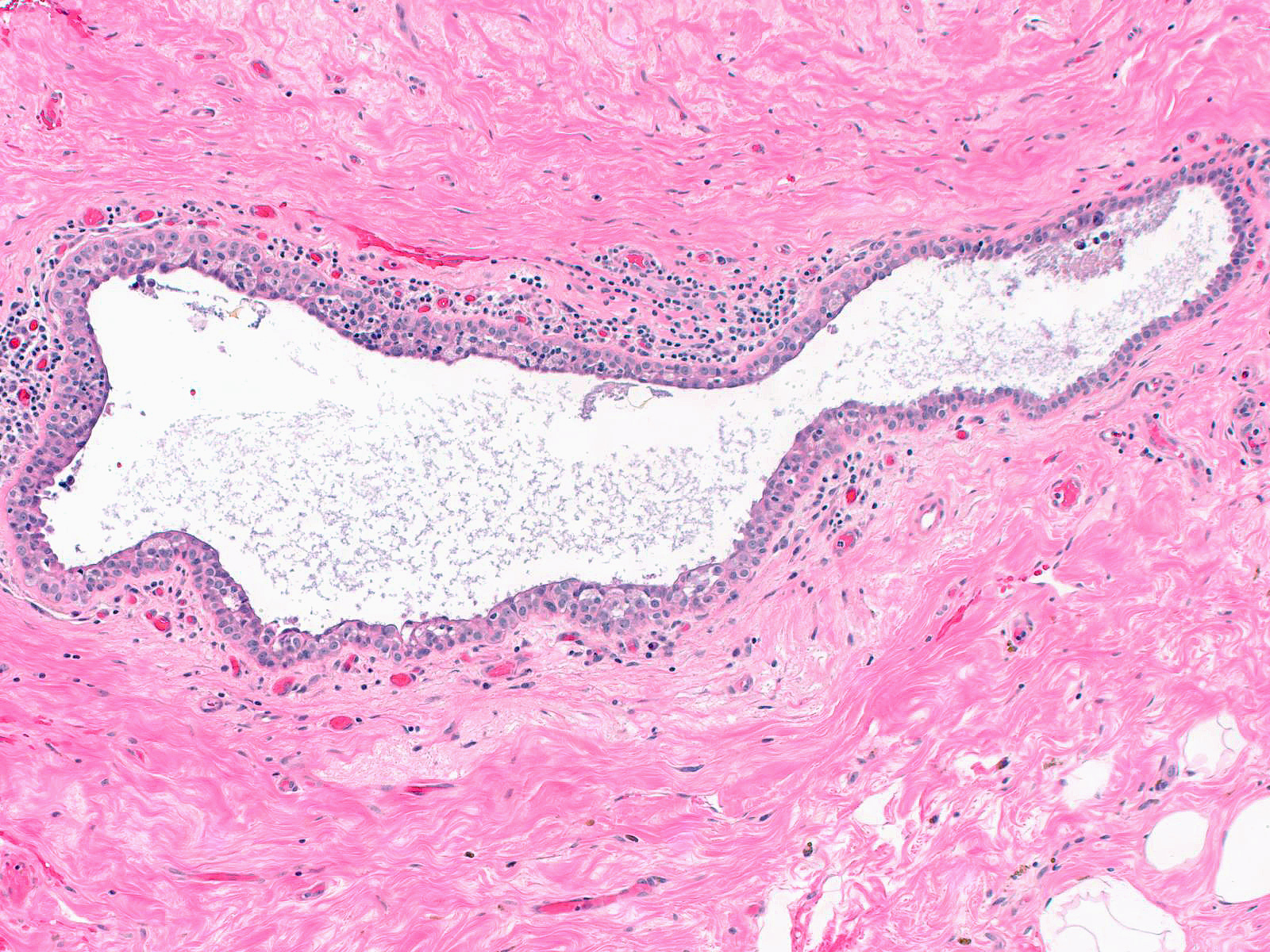 |
Nonspecialized stroma (also called extralobular stroma) surrounds the specialized stroma of ducts and ductules and encloses entire lobules. The nonspecialized stroma provides the structure of the breast and supports the function of the glandular tissue. Nonspecialized stroma consists of bundles of dense, eosinophilic collagen with intermixed large and small blood vessels, lymphatic vessels, fibroblasts, and myofibroblasts. Anteriorly, the nonspecialized stroma blends with the fibrous tissue of the reticular dermis and posteriorly it blends with the prepectoral fascia. Fibrous bands extending anteriorly from the prepectoral fascia are referred to as Cooper's ligaments. The fibrous connective tissue of the nonspecialized stroma intermixes with adipose tissue.
| An anterior view of the fibrous tissue which supports the breast and envelopes the glandular network. The nipple is seen in the top center of the image. (Cooper, 1840) | 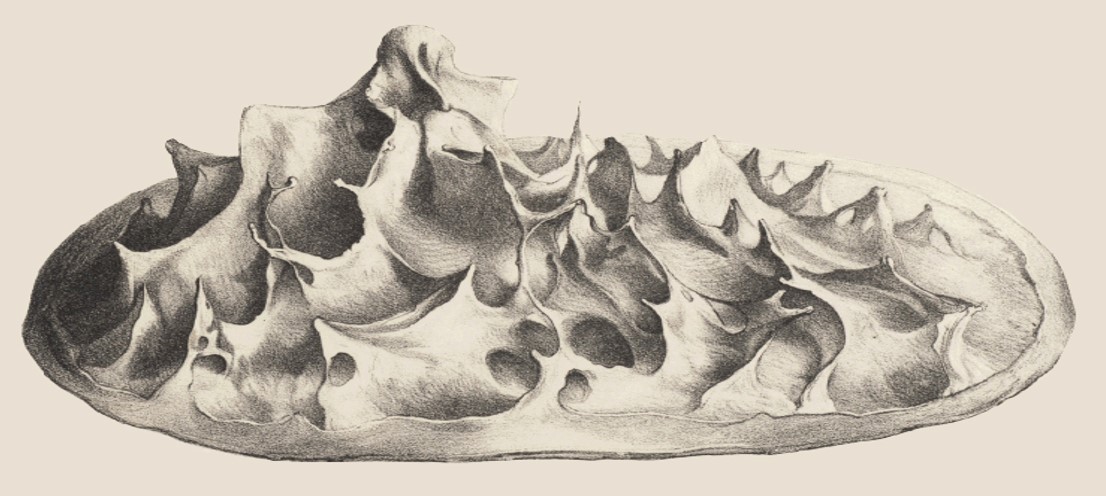 |
| Section through the nipple showing the glandular network and suspensory fibrous tissue. The fibrous tissue forms two partitions, anchoring the gland anteriorly to the skin and posteriorly to the pectoral muscle. (Cooper, 1840) | 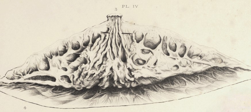 |
| During development, the primitive epidermis of the anterior chest invaginates into the mesoderm to create the basic structure of the breast's glandular component. Despite the complexity of the final structure, the breast maintains its layered compartments, which could be stretched out to reveal flat sheets that never intermix. The layers of the breast from most external to most internal are: (1) luminal epithelial cells; (2) myoepithelial cells; (3) basement membrane; (4) specialized stroma; (5) non-specialized stroma; and (6) fat. Mixing of non-juxtaposed layers likely represents a pathologic process. | 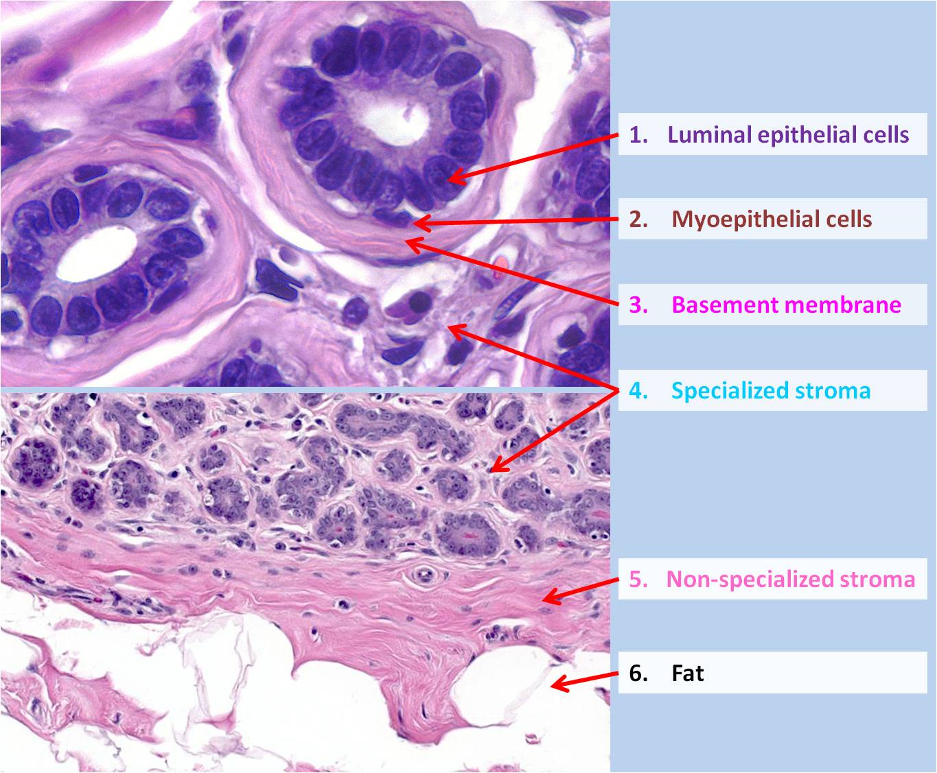 |
Lymphoid Tissue
The breast contains mucosa associated lymphoid tissue. T-cell aggregates occupy the specialized stroma around the walls of large ducts and occasionally infiltrates the overlying epithelium, which consequently appears hyperplastic. These T-cells probably serve to detect offending antigens. The specialized stroma of lobules also contains plasma cells and their precursors. The plasma cells produce the secretory IgA present in milk. Collections of B-cells may encircle small veins.
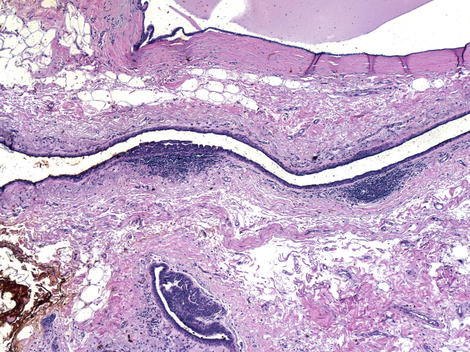 |
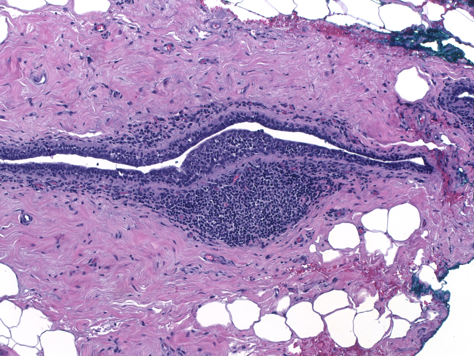 |
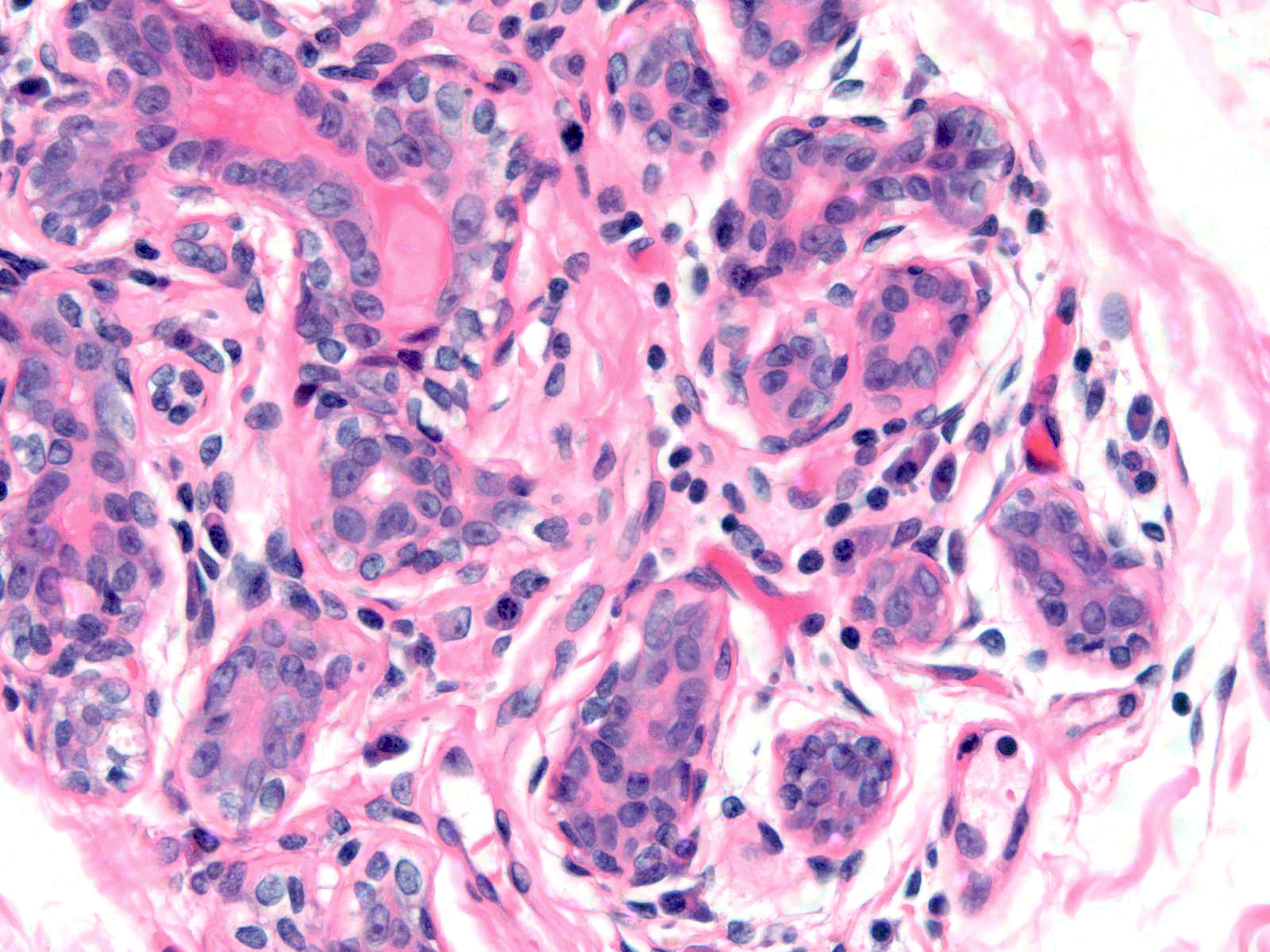 |
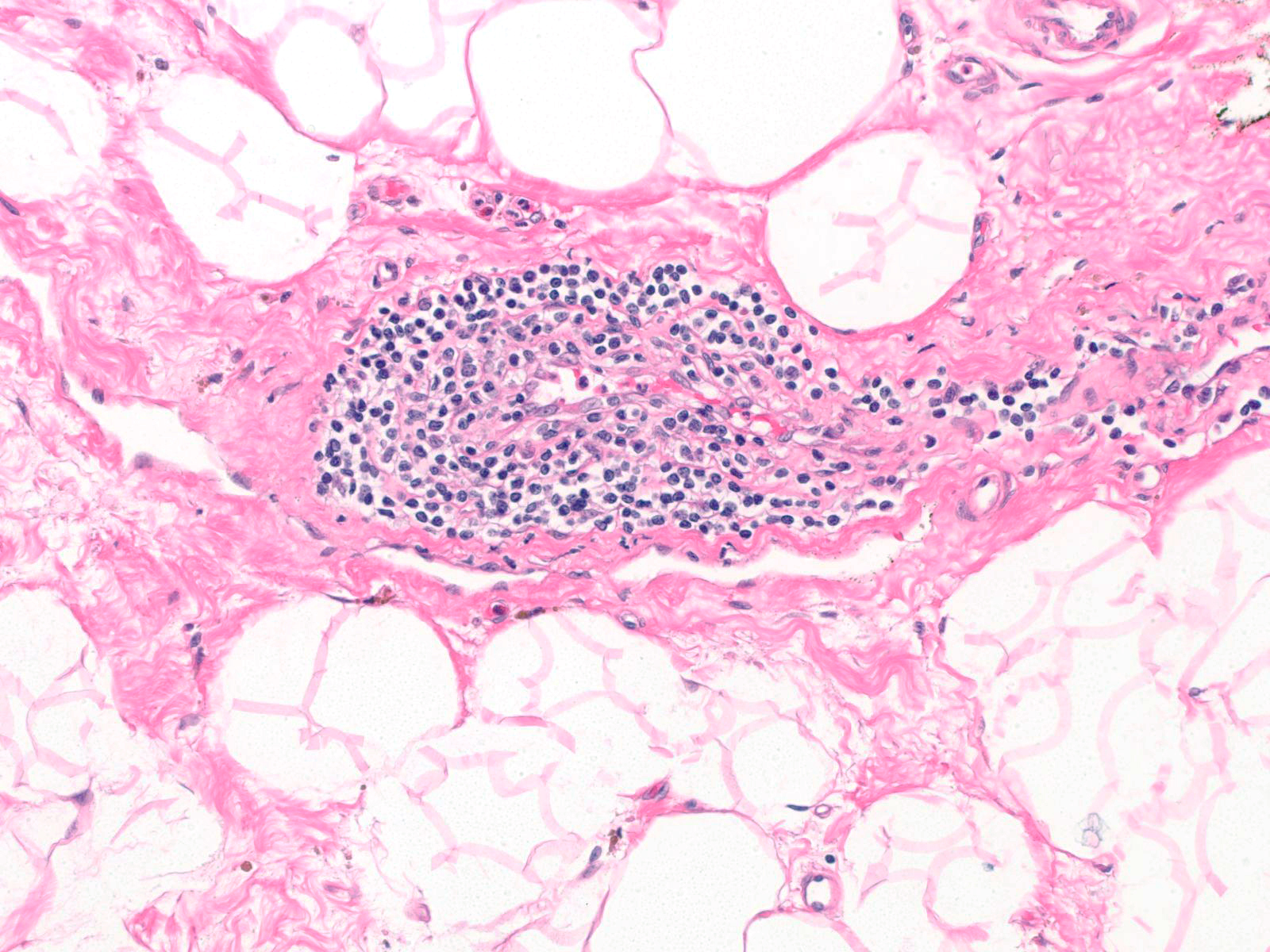 |
Skin
The skin covering the breast resembles the skin covering the remainder of the body. It consists of stratified squamous epithelium and the usual adnexal structures. Like other apocrine-gland-bearing skin, mammary epithelium contains Toker cells. These cells are found near the orifices of the lactiferous sinuses and other ducts that drain onto the skin of the nipple and areola. They sit in the basal region of the epithelium and occur singly and in small groups. Toker cells appear about the same size as their neighboring keratinocytes and they contain bland nuclei, small nucleoli, and pale, finely eosinophilic cytoplasm. Toker cells stain for CK7, but they do not stain for Her2. Toker cells may give rise to the malignant cells in certain cases of Paget disease of the breast. The lack of nuclear pleomorphism, the restriction to the basal aspect of the epidermis, and the absence of staining for HER2 help to distinguish Toker cells from the carcinoma cells of Paget disease.
| Toker cells are basally oriented, about the same size as keratinocytes, contain bland nuclei, and stain for keratin 7 but not Her2. | 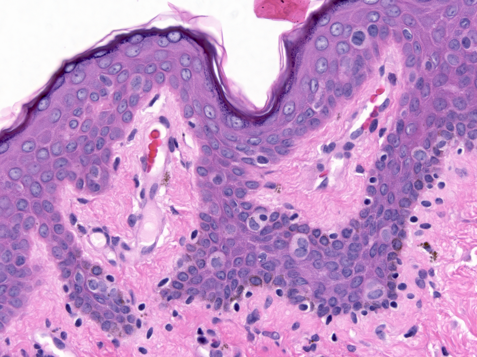 |
Quiz
<https://hub.partners.org/pathology/assessment/instructions?assessment_id=16607586" height="666" width="100%"></htmltag>