|
|
| (2 intermediate revisions by the same user not shown) |
| Line 23: |
Line 23: |
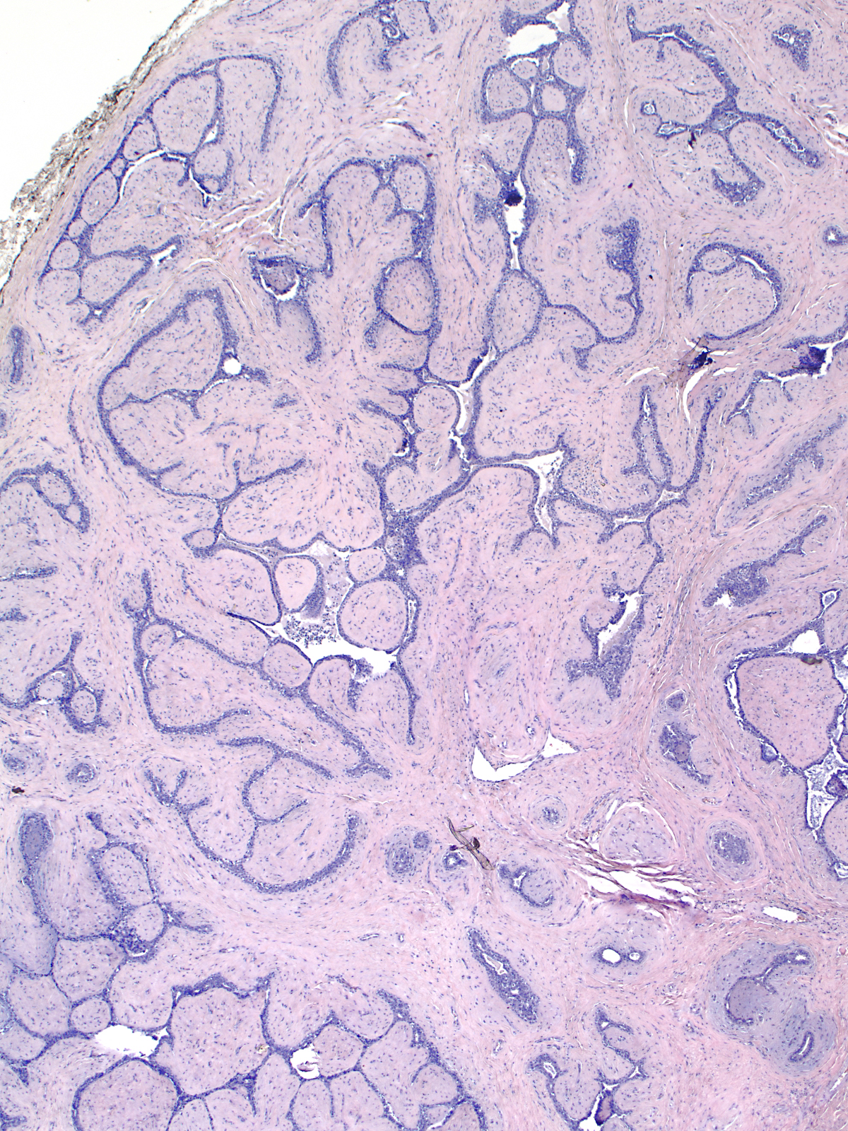
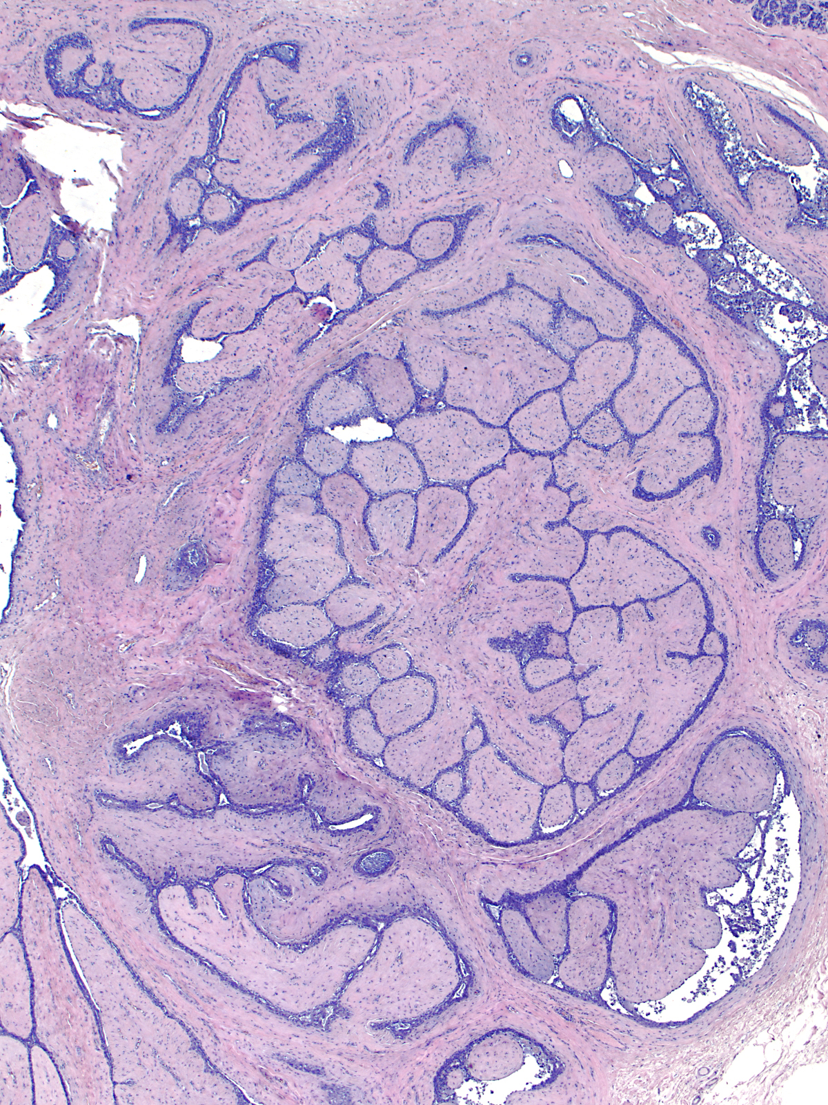
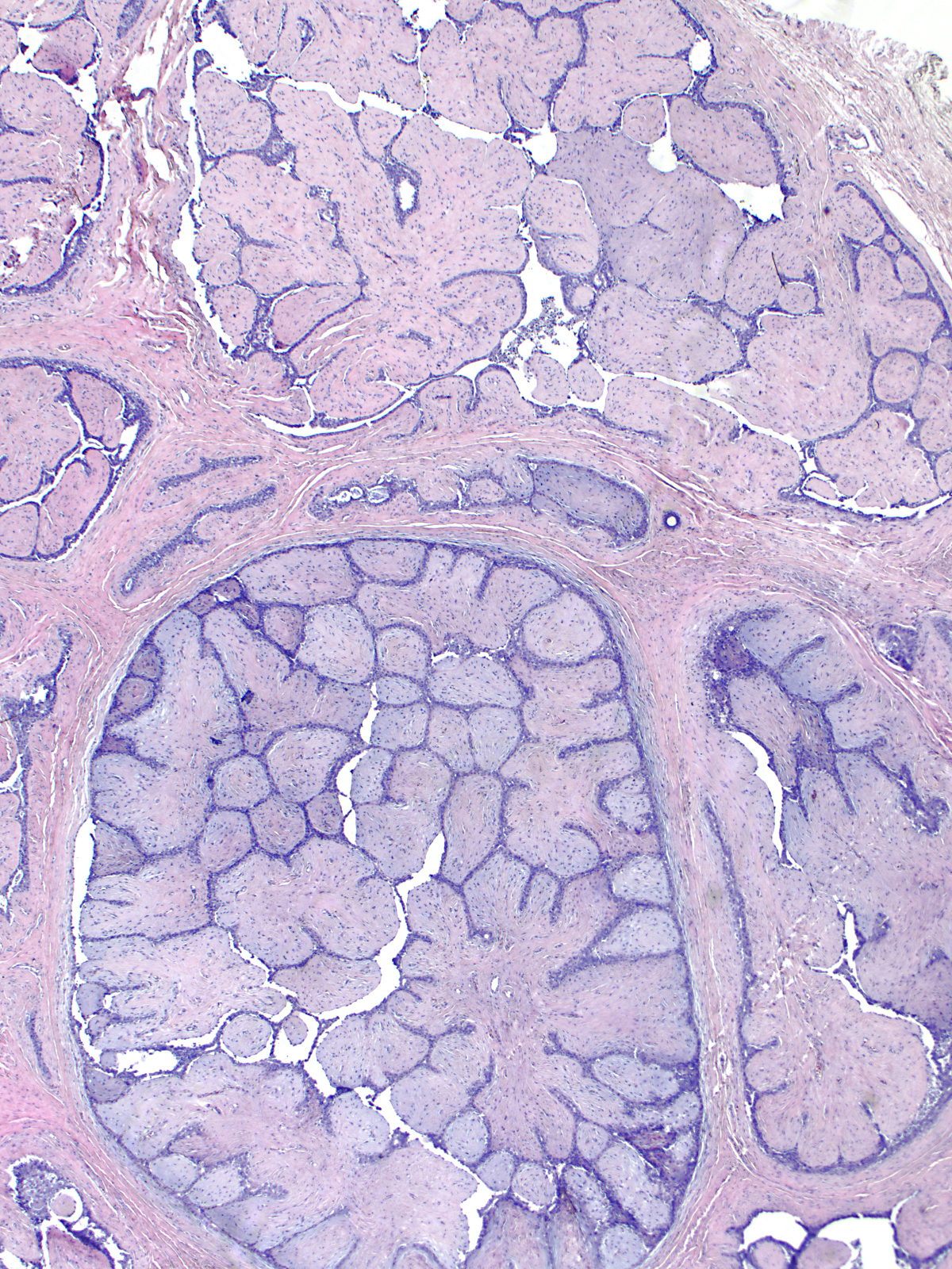
| | Although the histological features of fibroadenomas vary depending on the age and the hormonal status of the patient, the features do not differ much from one region to the next within a single tumor. The cellularity of the stroma, the nature of the extracellular matrix, the configuration of the glands, and the ratio of glands to stroma look similar throughout the mass. Below are three widely spaced foci from a single fibroadenoma.|||3|}} | | Although the histological features of fibroadenomas vary depending on the age and the hormonal status of the patient, the features do not differ much from one region to the next within a single tumor. The cellularity of the stroma, the nature of the extracellular matrix, the configuration of the glands, and the ratio of glands to stroma look similar throughout the mass. Below are three widely spaced foci from a single fibroadenoma.|||3|}} |
| | {{img3|FBA_1.jpg|FBA_2.jpg|FBA_3.jpg|}} | | {{img3|FBA_1.jpg|FBA_2.jpg|FBA_3.jpg|}} |
| − | == Quiz ==
| + | {{:mgh:breast-footer}} |
| − | <bootstrap_collapse text="Start Quiz"><p><htmltag tagname="iframe" src="https://hub.partners.org/pathology/assessment/assessment?assessment_id=16582314" height="666" width="100%"></htmltag>
| |
| − | </p></bootstrap_collapse>
| |
| − | <hr>
| |
| − | <categorytree mode="pages">Breast</categorytree>
| |
| − | [[Category:Breast • Weeks 2 and 3 • Diagnostic Entities]]
| |
Latest revision as of 18:09, July 13, 2020
Introduction
|
Definition: Fibroadenoma (FBA) is a fibroma that originates from fibroblasts of the specialized stroma. The entity belongs to the family of fibroepithelial lesions.
Clinical Significance: Fibroadenomas can come to clinical attention in women of any age, but they most commonly present in young women. Palpable fibroadenomas typically arise in women in their second and third decades. Radiological screening detects fibroadenomas in women of all screened ages, but fibroadenomas almost never arise in women beyond the age of forty years. Fibroadenomas form well-defined, mobile, rubbery, painless masses. They usually occur singly, but patients can develop multiple, successive, or bilateral tumors. The nodules usually span no more than a few centimeters and, once detected, they do not enlarge. During pregnancy, fibroadenomas can grow to massive sizes and undergo spontaneous infarction.
The well-defined borders of fibroadenomas allow surgeons to enucleate the tumors. They do not recur, although patients may develop additional fibroadenomas. In rare instances, fibroadenomas give rise to phyllodes tumors.
Gross Findings: Fibroadenomas appear as well defined, rubbery masses composed of lobules of pale yellow, white, or grey tissue, which bulges above the cut surface.
Microscopic Findings: The tumor consists of a nodular proliferation of fibroblasts, which expand the specialized stromal compartment and entrap and distort pre-existing ducts and lobules.
Differential Diagnosis: Hamartoma (malformed architecture), phyllodes tumor (stromal atypia), myxoid fibroadenoma (no stromal proliferation)
Discussion: Fibroadenomas have a consistent structure and uniform histologic characteristics, which change with the patient's age and hormonal status.
|
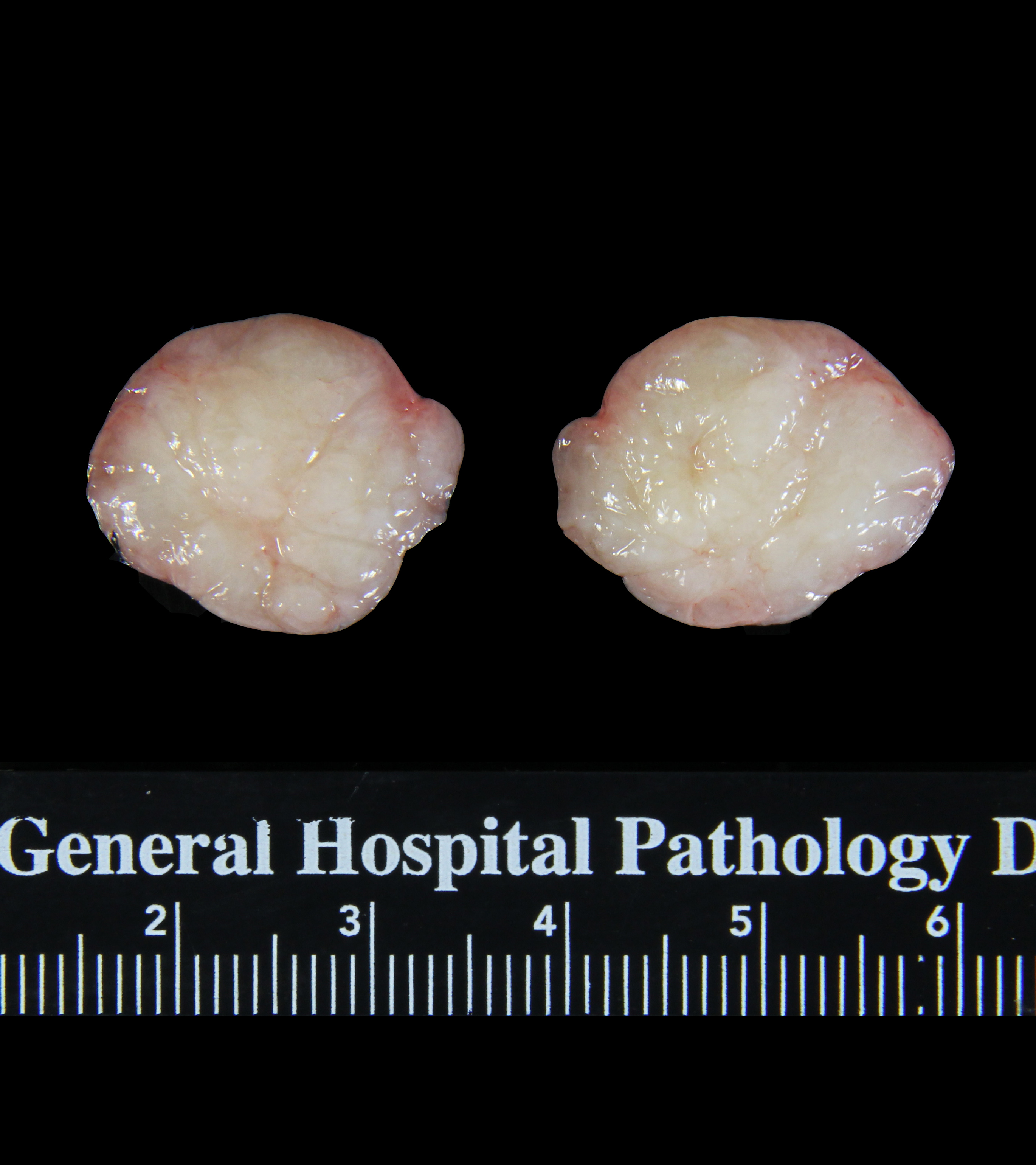 Grossly, FBAs appear as well-defined, rubbery masses that bulge above the cut surface. Grossly, FBAs appear as well-defined, rubbery masses that bulge above the cut surface. |
Origin of FBA
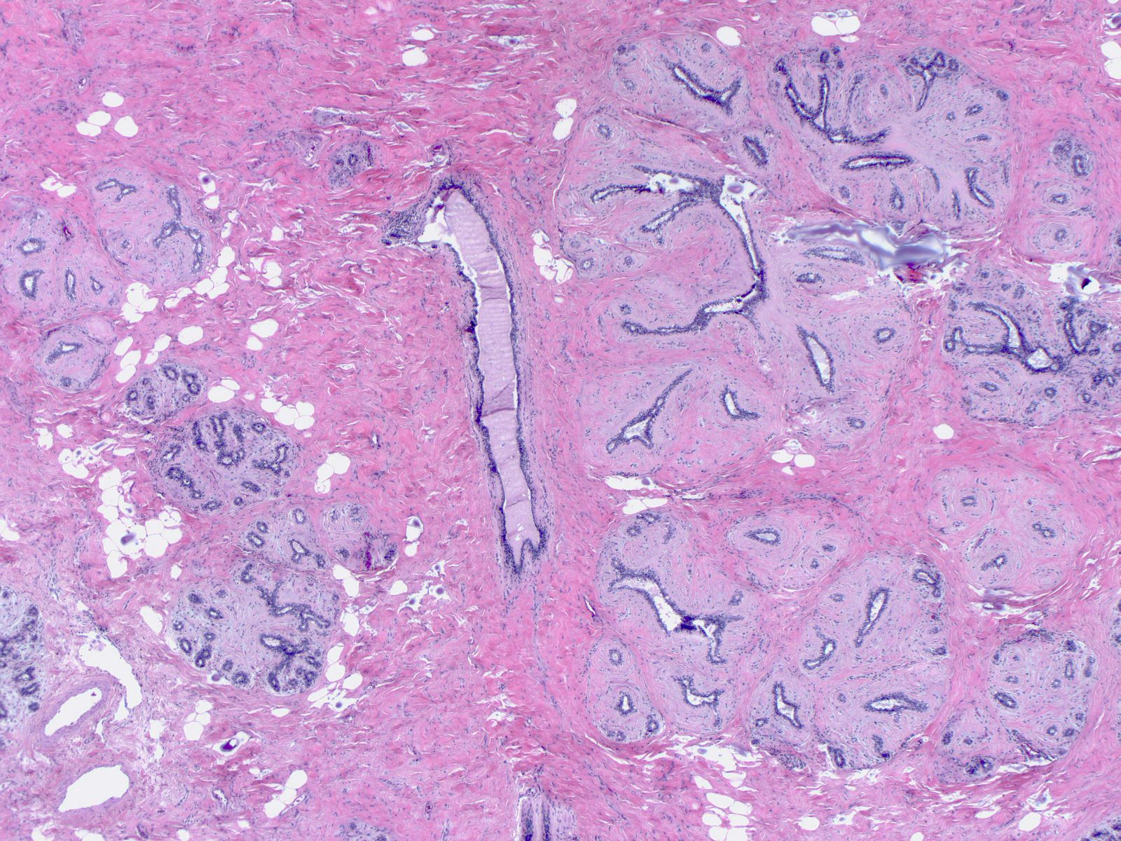
| Most fibroadenomas arise in the peripheral regions of the mammary ductal tree. As the fibroblasts multiply, they expand the specialized stromal compartment and distort the associated terminal ductules and acini (right side of image). |
 |
|
|
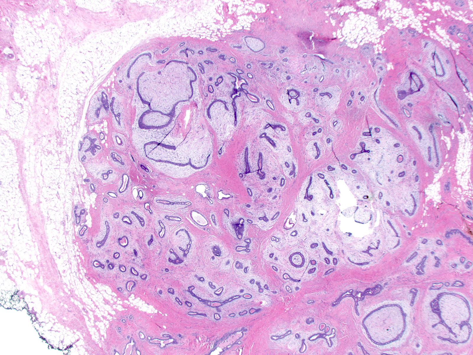
| With continued multiplication of the neoplastic stromal cells, the involved lobules and small ducts coalesce and form a mass. The growing mass extends along the glandular tree, incorporating more and more glandular tissue, until it has reached a size of a few centimeters, when growth usually stops. |
 |
|
|
Structure of FBA
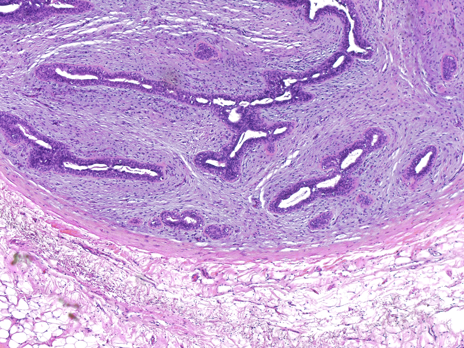
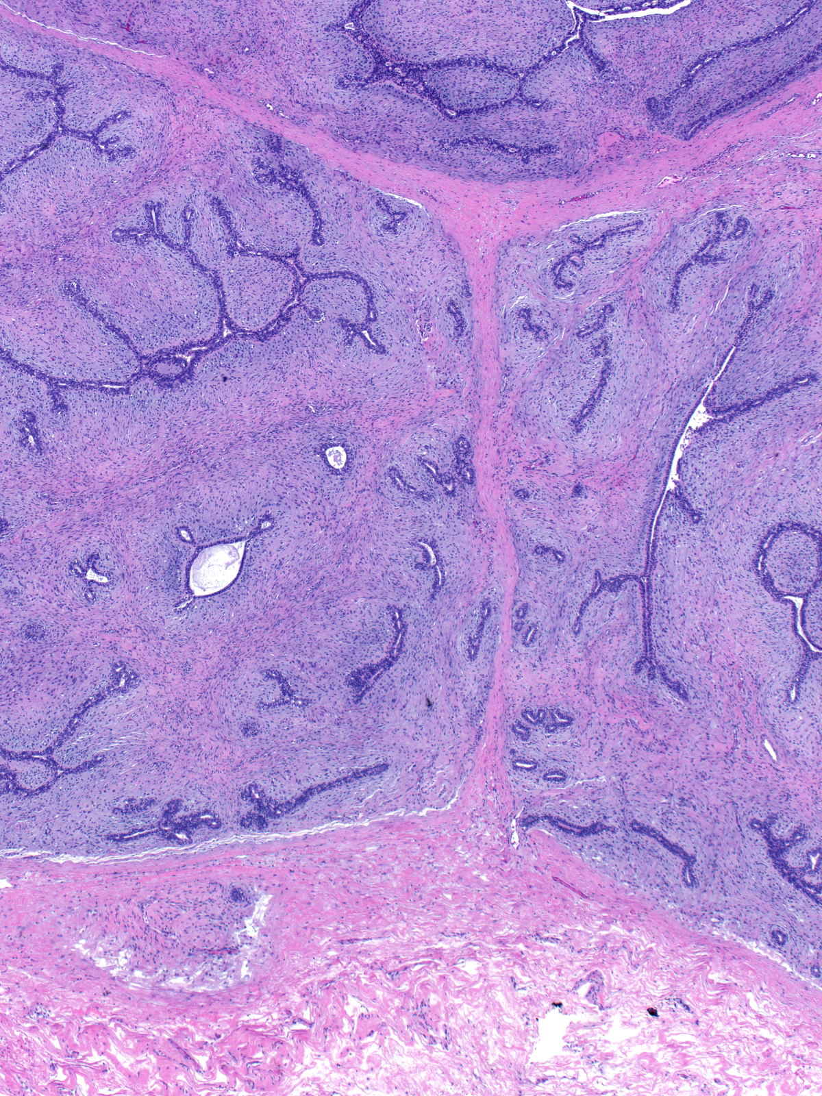
| Fibroadenomas display uniform histological characteristics. A zone of nonspecialized stroma forms a rim around the mass. The proliferating stroma cells do not penetrate into the surrounding tissue, nor does fat become incorporated into the tumor. |
 |
|
|
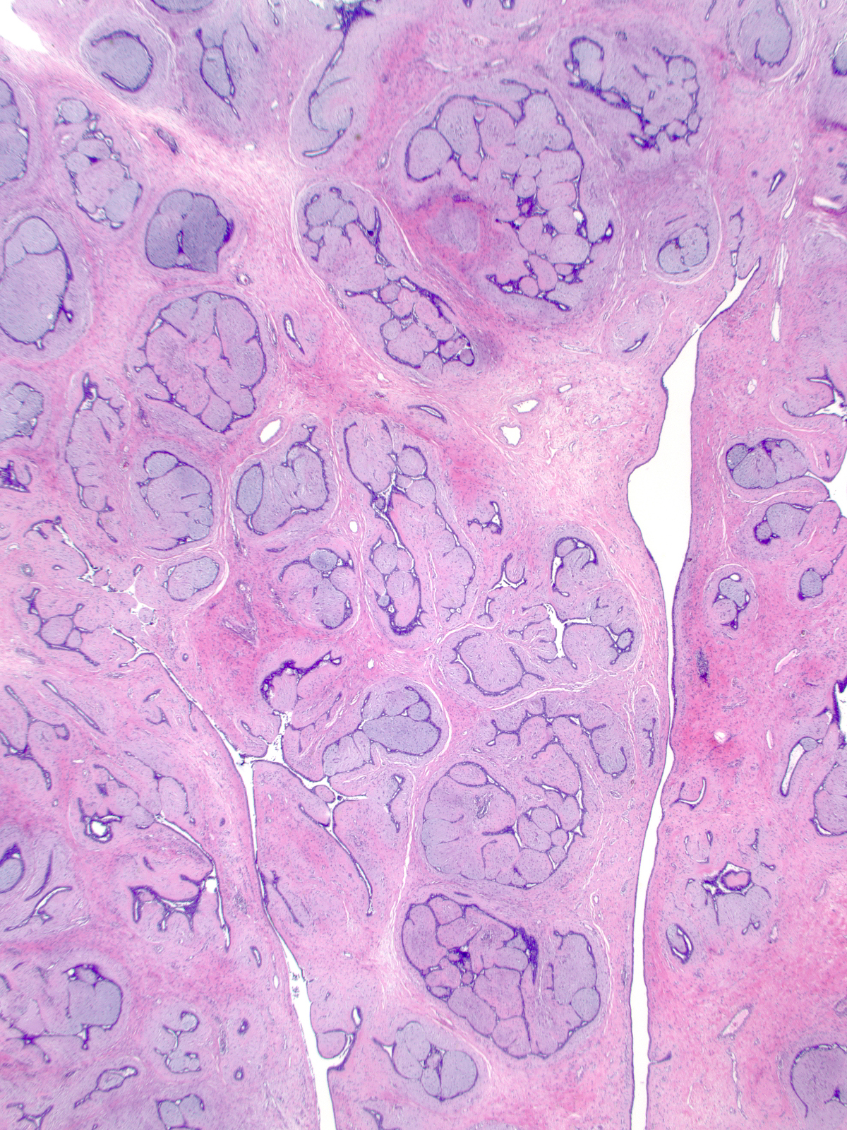
| Bands of nonspecialized stroma extend into the mass and subdivide it into units. Each unit consists of compressed canaliculi surrounded by sheaths of neoplastic specialized stroma. |
 |
 |
|
|
|
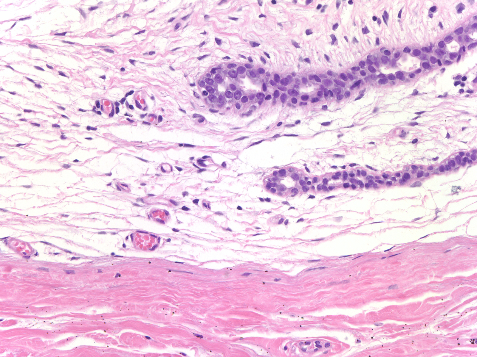
| The sheaths of specialized stroma at the periphery of the fibroadenoma appear thinner and less developed than those in the center. |
 |
|
|
Stroma of FBA
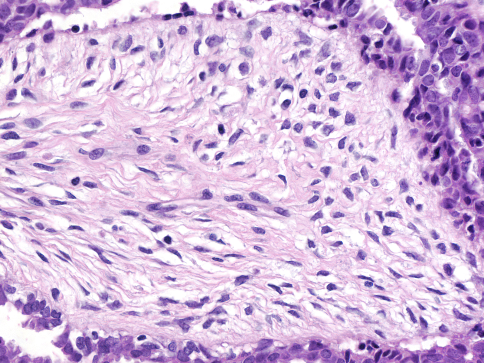
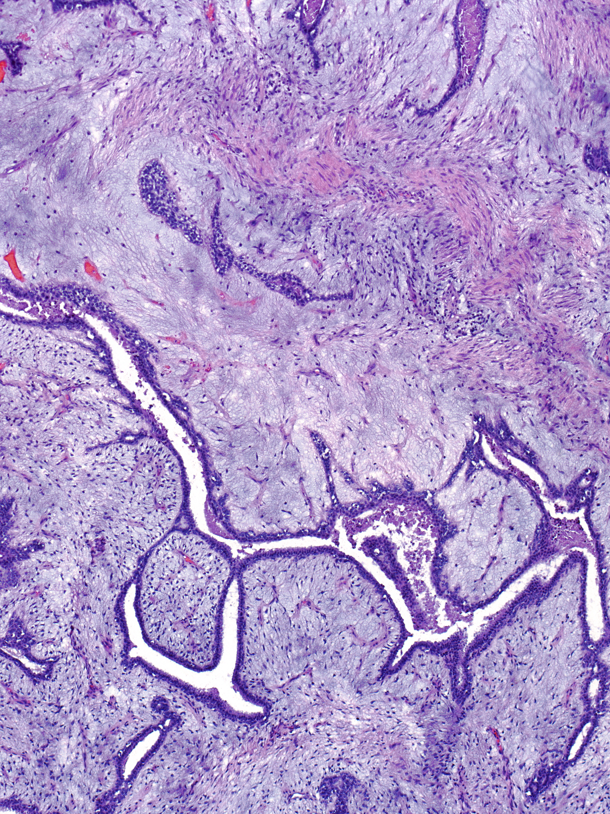
| The neoplastic specialized stroma consists of fibroblasts with small bland nuclei and inconspicuous nucleoli embedded in extracellular matrix. |
 |
|
|
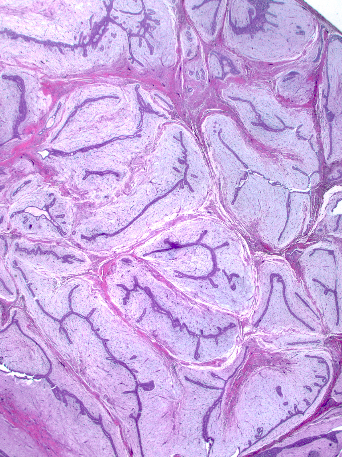
| Pale blue myxoid material makes up the matrix of fibroadenomas in young women. |
 |
 |
|
|
|
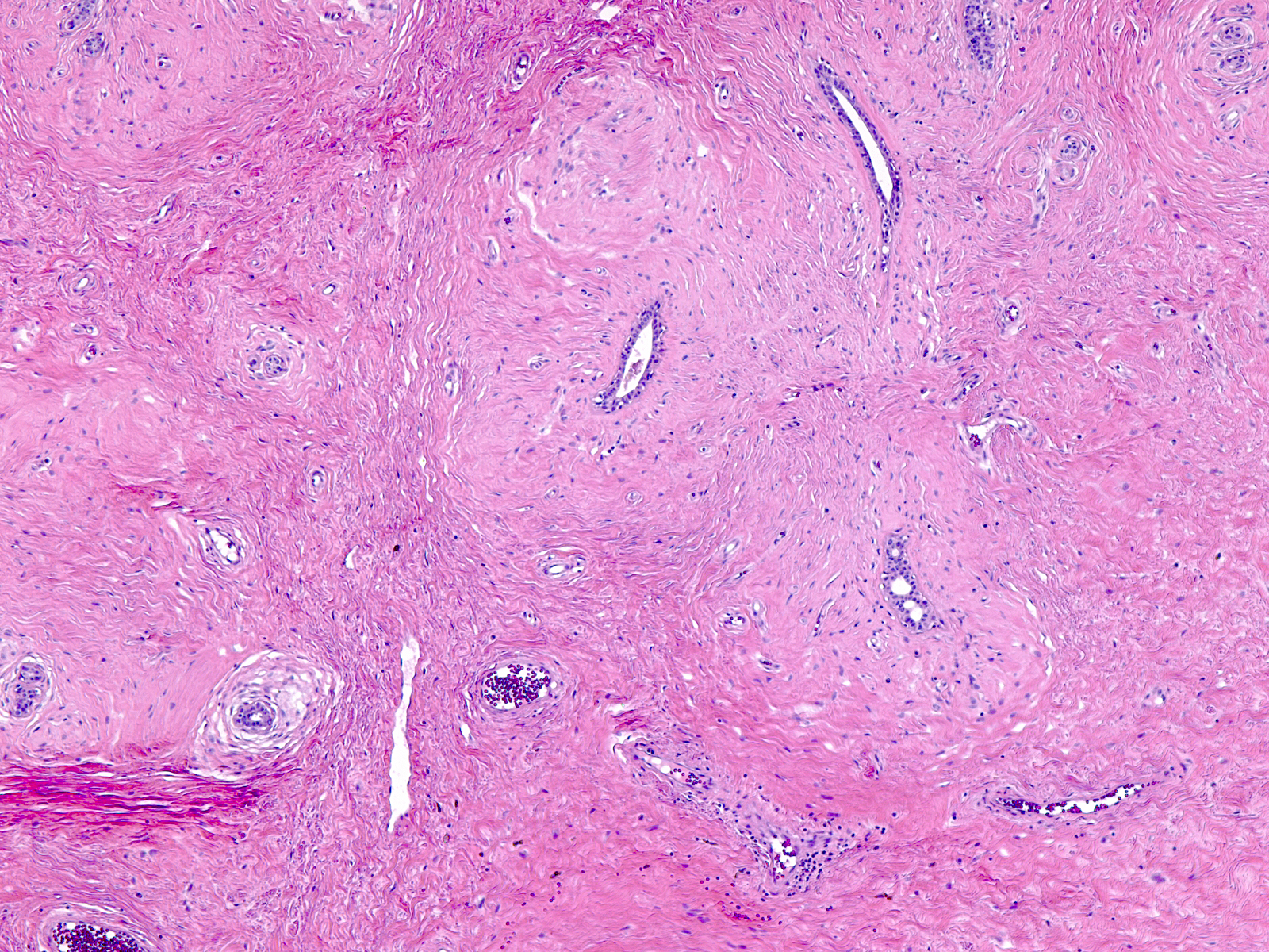
| With the passage of time, collagen replaces the myxoid material (hyalinization) and the specialized stroma comes to resemble the nonspecialized stromal bands. Attention to the hue and the orientation of the collagen bundles allows you to distinguish the two types of stroma in an older hyalinized fibroadenoma. |
 |
|
|
Glandular tissue of FBA
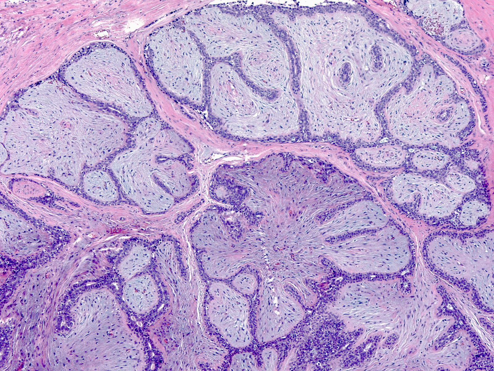
| The glandular tissue of fibroadenomas consists of slender, arching canaliculi with narrow, often inapparent, lumens. The canaliculi represent distorted pre-existing ductules and acini. |
 |
|
|
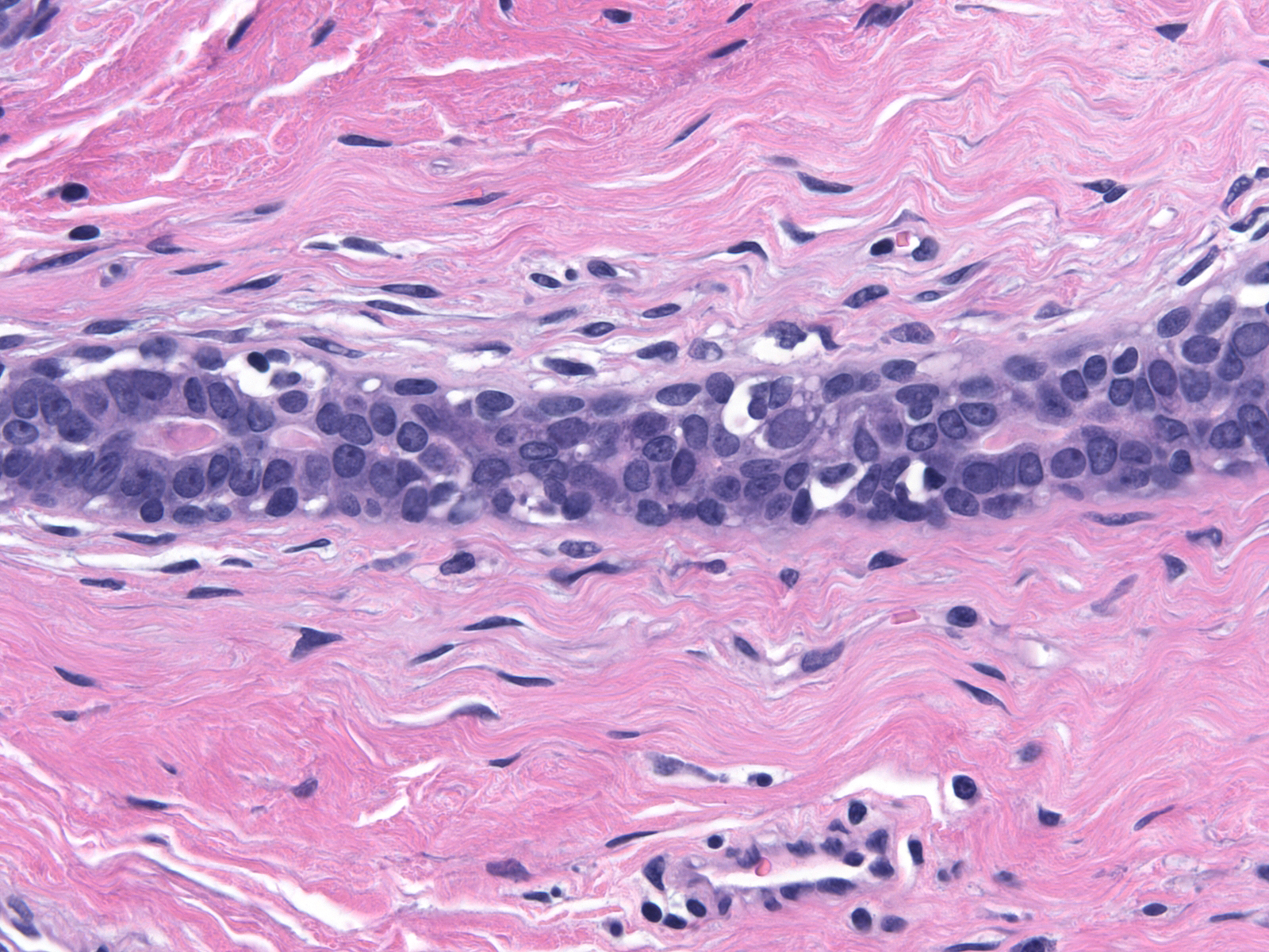
| The epithelium of the canaliculi appears inactive and consists of cuboidal to low columnar luminal cells with bland nuclei, small nucleoli, and small amounts of cytoplasm. The myoepithelial cells also appear small. |
 |
|
|
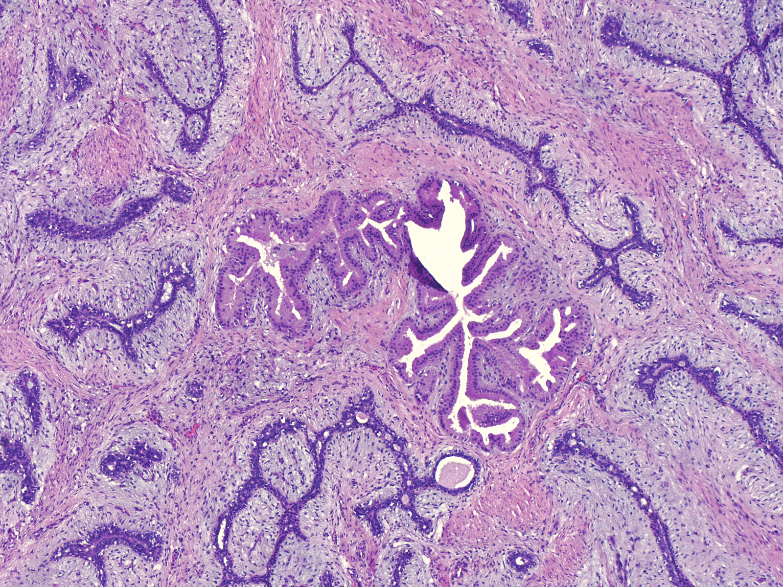
| Very rarely, apocrine metaplasia can affect the glandular tissue in fibroadenomas. |
 |
|
|
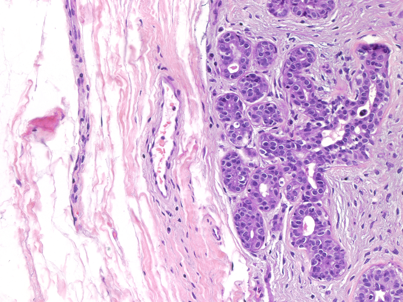
| The glands at the edge of a fibroadenoma often appears less distorted than the glands in the center of the mass, a hint that the compressed canaliculi were once normal ductules and acini. |
 |
|
|
Uniformity of FBA
Although the histological features of fibroadenomas vary depending on the age and the hormonal status of the patient, the features do not differ much from one region to the next within a single tumor. The cellularity of the stroma, the nature of the extracellular matrix, the configuration of the glands, and the ratio of glands to stroma look similar throughout the mass. Below are three widely spaced foci from a single fibroadenoma.
Breast Pathology


